Achilles tendinitis is a common condition that occurs when the large tendon that runs down the back of your lower leg becomes irritated and inflamed.
The Achilles tendon is the largest tendon in the body. It connects your calf muscles to your heel bone and is used when you walk, run, climb stairs, jump, and stand on your tip toes. Although the Achilles tendon can withstand great stresses from running and jumping, it is also prone to tendinitis, a condition associated with overuse and degeneration.
What Is the Achilles Tendon?
A tendon is a band of tissue that connects a muscle to a bone. The Achilles tendon runs down the back of the lower leg and connects the calf muscle to the heel bone. Also called the heel cord, the Achilles tendon facilitates walking by helping to raise the heel off the ground.
What Is an Achilles Tendon Rupture?
An Achilles tendon rupture is a complete or partial tear that occurs when the tendon is stretched beyond its capacity. Forceful jumping or pivoting, or sudden accelerations of running, can overstretch the tendon and cause a tear. An injury to the tendon can also result from falling or tripping.
Achilles tendon ruptures are most often seen in “weekend warriors”—typically, middle-aged people participating in sports in their spare time. Less commonly, illness or medications, such as steroids or certain antibiotics, may weaken the tendon and contribute to ruptures.
Signs & Symptoms
A person with a ruptured Achilles tendon may experience one or more of the following:
- Sudden pain (which feels like a kick or a stab) in the back of the ankle or calf—often subsiding into a dull ache
- A popping or snapping sensation
- Swelling on the back of the leg between the heel and the calf
- Difficulty walking (especially upstairs or uphill) and difficulty rising up on the toes
These symptoms require prompt medical attention to prevent further damage. Until the patient is able to see a doctor, the RICE method should be used. This involves:
- Rest. Stay off the injured foot and ankle, since walking can cause pain or further damage.
- Ice. Apply a bag of ice covered with a thin towel to reduce swelling and pain. Do not put ice directly against the skin.
- Compression. Wrap the foot and ankle in an elastic bandage to prevent further swelling.
- Elevation. Keep the leg elevated to reduce the swelling. It should be even with or slightly above heart level.
Diagnosis
In diagnosing an Achilles tendon rupture, the foot and ankle surgeon will ask questions about how and when the injury occurred and whether the patient has previously injured the tendon or experienced similar symptoms. The surgeon will examine the foot and ankle, feeling for a defect in the tendon that suggests a tear. Range of motion and muscle strength will be evaluated and compared to the uninjured foot and ankle. If the Achilles tendon is ruptured, the patient will have less strength in pushing down (as on a gas pedal) and will have difficulty rising on the toes.
The diagnosis of an Achilles tendon rupture is typically straightforward and can be made through this type of examination. In some cases, however, the surgeon may order an MRI or other advanced imaging tests.
Treatment
Treatment options for an Achilles tendon rupture include surgical and nonsurgical approaches. The decision of whether to proceed with surgery or nonsurgical treatment is based on the severity of the rupture and the patient’s health status and activity level.
Nonsurgical Treatment
Nonsurgical treatment, which is generally associated with a higher rate of rerupture, is selected for minor ruptures, less active patients and those with medical conditions that prevent them from undergoing surgery. Nonsurgical treatment involves use of a cast, walking boot or brace to restrict motion and allow the torn tendon to heal.
Surgery
Surgery offers important potential benefits. Besides decreasing the likelihood of rerupturing the Achilles tendon, surgery often increases the patient’s push-off strength and improves muscle function and movement of the ankle.
Various surgical techniques are available to repair the rupture. The surgeon will select the procedure best suited to the patient.
Following surgery, the foot and ankle are initially immobilized in a cast or walking boot. The surgeon will determine when the patient can begin weightbearing.
Complications such as incision-healing difficulties, rerupture of the tendon or nerve pain can arise after surgery.
Physical Therapy
Whether an Achilles tendon rupture is treated surgically or nonsurgically, physical therapy is an important component of the healing process. Physical therapy involves exercises that strengthen the muscles and improve range of motion in the foot and ankle.
Flat feet, also known as fallen arches, are a postural deformity in which the bottom of your foot makes full or nearly entire contact with the ground. Flat feet impact millions of people worldwide and, in most cases, do not cause problems. However, more severe forms of flat feet can cause joint discomfort and stiffness, particularly in the ankles and knees

Tendons connect muscles to bones and stretch across joints, enabling you to bend that joint. One of the most important tendons in the lower leg is the posterior tibial tendon. This tendon starts in the calf, stretches down behind the inside of the ankle and attaches to bones in the middle of the foot.
The posterior tibial tendon helps hold your arch up and provides support as you step off on your toes when walking. If this tendon becomes inflamed, over-stretched or torn, you may experience pain on the inner ankle and gradually lose the inner arch on the bottom of your foot, leading to flatfoot.
Flatfoot Signs & Symptoms
- Pain and swelling on the inside of the ankle
- Loss of the arch and the development of a flatfoot
- Weakness and an inability to stand on the toes
- Tenderness over the inside of the ankle when active
Risk Factors
- Women over 50 years of age
- Obesity
- Diabetes
- Hypertension
- Previous surgery or trauma (ankle fracture)
- Inflammatory diseases such as Rheumatoid Arthritis
Diagnosis
The diagnosis is based on both a history and a physical examination. Your physician may ask you to stand on your bare feet facing away from him/her to view how your foot functions. As the condition progresses, the front of the affected foot will start to slide to the outside. From behind, it will look as though you have “too many toes” showing. You may also be asked to stand on your toes or to do a single heel rise: stand with your hands on the wall, lift the unaffected foot off the ground, and raise up on the toes of the other foot. Normally, the heel will rotate inward; the absence of this sign indicates posterior tibial tendon dysfunction. Your doctor may request X-rays or a magnetic resonance image (MRI) of the foot.
Treatment Options for Flatfoot
Without treatment, the flatfoot that develops from posterior tibial tendon dysfunction eventually becomes rigid. Arthritis develops in the hindfoot. Pain increases and spreads to the outer side of the ankle. The way you walk may be affected and wearing shoes may be difficult.
The treatment your doctor recommends will depend on how far the condition has progressed. In the early stages, posterior tibial tendon dysfunction can be treated with rest, non-steroidal anti-inflammatory drugs such as aspirin or ibuprofen, and immobilization of the foot for 6 to 8 weeks with a rigid below-knee cast or boot to prevent overuse. After the cast is removed, shoe inserts such as a heel wedge or arch support may be helpful. If the condition is advanced, your doctor may recommend that you use a custom-made ankle-foot orthosis or support.
If conservative treatments don’t work, your doctor may recommend surgery. Several procedures can be used to treat posterior tibial tendon dysfunction; often more than one procedure is performed at the same time. Your doctor will recommend a specific course of treatment based on your individual case.
Ankle sprains are caused by an unnatural twisting or force on the ankle bones of the foot, which may result in excessive stretching or tearing of one or more ligaments on the outside of the ankle. The severity of the sprain can impact the degree of damage as well as the type and duration of treatment. If not properly treated, ankle sprains may develop into long-term problems.
Primary symptoms of ankle sprains are pain following a twist or injury, swelling, and bruising.
Treatment includes resting and elevating the ankle and applying ice to reduce swelling. Compressive bandages also may be used to immobilize and support the injury during healing. Serious ankle sprains, particularly among competitive athletes, may require surgery to repair and tighten the damaged ligaments.
To prevent ankle sprains, try to maintain strength, balance, and flexibility in the foot and ankle through exercising, stretching, and wearing well-fitted shoes.
A chronic infection caused by various types of fungus, Athlete’s foot is often spread in places where people go barefoot such as public showers or swimming pools. The condition ranges from mild scaling and itching to painful inflammation and blisters. It usually starts between the toes or on the arch and may spread to the bottom and sides of the foot.
General Treatments
Depending on the type of infection you have, various kinds of medication may be used in treating your fungal problem. Successful treatment usually involves a combination of medication and self-care.
Hallux rigidus is a condition that causes stiffness and pain in the big toe joint. It is a form of osteoarthritis that affects the joint at the base of the big toe (the metatarsophalangeal joint). The condition is characterized by a loss of flexibility and range of motion in the big toe, as well as pain and swelling in the joint.
The severity of hallux rigidus can vary, with some people experiencing mild symptoms that only occur when they perform certain activities, while others may have more severe symptoms that affect their daily activities. The condition is more common in people over the age of 40, and it is more common in men than in women.
Hallux rigidus is usually caused by wear and tear on the joint, which can be exacerbated by activities that put a lot of stress on the joint, such as walking or running for long periods of time, or participating in high-impact sports. Other factors that can contribute to the development of hallux rigidus include foot deformities, previous injury to the joint, and certain medical conditions, such as rheumatoid arthritis.
Treatment for hallux rigidus usually involves a combination of conservative measures, such as rest, ice, and physical therapy, and may also include medications to reduce inflammation and pain. In more severe cases, surgery may be necessary to repair or reconstruct the joint
A broken ankle is also known as an ankle “fracture.” This means that one or more of the bones that make up the ankle joint are broken.
A fractured ankle can range from a simple break in one bone, which may not stop you from walking, to several fractures, which forces your ankle out of place and may require that you not put weight on it for a few months.
Simply put, the more bones that are broken, the more unstable the ankle becomes. There may be ligaments damaged as well. The ligaments of the ankle hold the ankle bones and joint in position.
Cause
- Twisting or rotating your ankle
- Rolling your ankle
- Tripping or falling
- Impact during a car accident
Symptoms
Because a severe ankle sprain can feel the same as a broken ankle, every ankle injury should be evaluated by a physician.
Common symptoms for a broken ankle include:
- Immediate and severe pain
- Swelling
- Bruising
- Tender to touch
- Cannot put any weight on the injured foot
- Deformity (“out of place”), particularly if the ankle joint is dislocated as well
Recovery
Because there is such a wide range of injuries, there is also a wide range of how people heal after their injury. It takes at least 6 weeks for the broken bones to heal. It may take longer for the involved ligaments and tendons to heal.
As mentioned above, your doctor will most likely monitor the bone healing with repeated x-rays. This is typically done more often during the first 6 weeks if surgery is not chosen.
Pain Management
Pain after an injury or surgery is a natural part of the healing process. Your doctor and nurses will work to reduce your pain, which can help you recover faster.
Medications are often prescribed for short-term pain relief after surgery or an injury. Many types of medicines are available to help manage pain, including opioids, non-steroidal anti-inflammatory drugs (NSAIDs), and local anesthetics. Your doctor may use a combination of these medications to improve pain relief, as well as minimize the need for opioids.
Be aware that although opioids help relieve pain after surgery or an injury, they are a narcotic and can be addictive. It is important to use opioids only as directed by your doctor. As soon as your pain begins to improve, stop taking opioids. Talk to your doctor if your pain has not begun to improve within a few days of your treatment.
Rehabilitation
Rehabilitation is very important regardless of how an ankle fracture is treated.
When your physician allows you to start moving your ankle, physical therapy and home exercise programs are very important. Doing your exercises regularly is key.
Eventually, you will also start doing strengthening exercises. It may take several months for the muscles around your ankle to get strong enough for you to walk without a limp and to return to your regular activities.
Again, exercises only make a difference if you actually do them.
Weightbearing
Your specific fracture determines when you can start putting weight on your ankle. Your physician will allow you to start putting weight on your ankle when he or she feels your injury is stable enough to do so.
It is very important to not put weight on your ankle until your physician says you can. If you put weight on the injured ankle too early, the fracture fragments may move or your surgery may fail and you may have to start over.
Supports
It is very common to have several different kinds of things to wear on the injured ankle, depending on the injury.
Initially, most ankle fractures are placed in a splint to protect your ankle and allow for the swelling to go down. After that, you may be put into a cast or removable brace.
Even after the fracture has healed, your physician may recommend wearing an ankle brace for several months while you are doing sporting activities.
Complications
People who smoke, have diabetes, or are elderly are at a higher risk for complications after surgery, including problems with wound healing. This is because it may take longer for their bones to heal.
Nonsurgical Treatment
Without surgery, there is a risk that the fracture will move out of place before it heals. This is why it is important to follow up with your physician as scheduled.
If the fracture fragments do move out of place and the bones heal in that position, it is called a “malunion.” Treatment for this is determined by how far out of place the bones are and how the stability of the ankle joint is affected.
If a malunion does occur or if your ankle becomes unstable after it heals, this can eventually lead to arthritis in your ankle.
Surgical Treatment
General surgical risks include:
- Infection
- Bleeding
- Pain
- Blood clots in your leg
- Damage to blood vessels, tendons, or nerves
Risks from the surgical treatment of ankle fractures include
- Difficulty with bone healing
- Arthritis
- Pain from the plates and screws that are used to fix fracture. Some patients choose to have them removed several months after their fracture heals
Outcomes
Although most people return to normal daily activities, except for sports, within 3 to 4 months, studies have shown that people can still be recovering up to 2 years after their ankle fractures. It may take several months for you to stop limping while you walk, and before you can return to sports at your previous competitive level. Most people return to driving within 9 to 12 weeks from the time they were injured.
A fracture of the calcaneus, or heel bone, can be a painful and disabling injury. This type of fracture commonly occurs during a high-energy event — such as a car crash or a fall from a ladder — when the heel is crushed under the weight of the body. When this occurs, the heel can widen, shorten, and become deformed.
Calcaneus fractures can be quite severe. Treatment often involves surgery to reconstruct the normal anatomy of the heel and restore mobility so that patients can return to normal activity. But even with appropriate treatment, some fractures may result in long-term complications, such as pain, swelling, loss of motion, and arthritis. Many patients with labor-intensive jobs are unable to return to their job after a calcaneus fracture.
Symptoms
Patients with calcaneus fractures usually experience:
- Pain
- Bruising
- Swelling
- Heel deformity
- Inability to put weight on the heel or walk
Nonsurgical Treatment
Your doctor may recommend nonsurgical treatment if the pieces of broken bone have not been displaced by the force of the injury.
Immobilization. A cast, splint, or brace will hold the bones in your foot in proper position while they heal. You may have to wear a cast for 6 to 8 weeks — or possibly longer. During this time, you will not be able to put any weight on your foot until the bone is completely healed.
Surgical Treatment
If the bones have shifted out of place (displaced), your doctor may recommend surgery.
Surgery to repair a calcaneus fracture can restore the normal shape of the bone but is sometimes associated with complications, such as wound healing problems, infection, and nerve damage.
Nonsurgical treatment of some fractures, however, can also lead to long-term complications, such as pain, arthritis, and a limp. Your doctor will review the details of your injury and talk with you about the risks and benefits of surgical versus nonsurgical treatment.
Timing of surgery. If the skin around your fracture has not been broken, your doctor may recommend waiting until swelling has gone down before having surgery. Elevating your leg and keeping it immobilized for several days will decrease swelling. It will also give injured skin a chance to recover. Waiting before the operation may improve your overall recovery from surgery and decrease your risk of infection.
Open fractures, however, expose the fracture site to the environment — increasing the risk of infection — and must be treated immediately. They require surgery to clean the wound and remove damaged tissue.
Early surgery may also be recommended for other fractures. Although uncommon, a piece of the calcaneus can be pulled off when the Achilles tendon splits away from the bone (avulsion). For this type of fracture, emergent surgery can decrease the risk of injury to the skin around the Achilles tendon.
Rehabilitation
Whether your treatment is surgical or nonsurgical, your rehabilitation will be very similar. The time it takes to return to daily activities will vary depending on the type and severity of the fracture and whether you have other injuries.
Some patients can begin weight-bearing activities a few weeks after injury or surgery; most will need to wait 3 months before putting weight on the heel. Some patients are able to begin partial weightbearing 6 to 10 weeks after injury or surgery.
Complications
Complications often occur with calcaneus fractures. Minor complications include:
- Small or temporary areas of delayed wound healing
- Nerve irritation around the incision
- Tendon irritation
- Joint stiffness
- Chronic pain
- Chronic swelling
Major complications include:
- Failure of the wound to heal
- Infection
- Posttraumatic arthritis(with or without surgery)
It is important to tell your doctor if you are a smoker. Smoking affects both bone and wound healing. With or without surgery, your bone may take longer to heal if you smoke.
Outcomes
If your injury is minor, such as a crack in the bone with little muscle damage, you may be able to resume normal activities 3 to 4 months after surgery. If your fracture is severe, however, it may take 1 to 2 years before recovery is complete.
Despite the best efforts of the doctor and patient, patients rarely regain normal foot and ankle motion after a severe fracture and do not typically resume their pre-injury level of function. A patient who is not very active might tolerate a foot that is not normal. On the other hand, a patient whose job or recreational activities require a lot of walking or climbing may require major lifestyle and career changes.
Toe and Metatarsal Fractures (Broken Toes)
Broken toes and broken metatarsal bones can be painful, significant injuries. The structure of the foot is complex, consisting of bones, muscles, tendons and other soft tissues. Of the 28 bones in the foot, 19 are toe bones (phalanges) and metatarsal bones (the long bones in the midfoot). Fractures of the toe and metatarsal bones are common and require evaluation by a specialist. A foot and ankle surgeon should be seen for proper diagnosis and treatment, even if initial treatment has been received in an emergency room.
What are Toe and Metatarsal Fractures?
A fracture is a break in the bone. Fractures can be divided into two categories: traumatic fractures and stress fractures.
Traumatic fractures (also called acute fractures) are caused by a direct blow or impact, such as seriously stubbing your toe. Traumatic fractures can be displaced or nondisplaced. If the fracture is displaced, the bone is broken in such a way that it has changed in position (malpositioned).
Signs and symptoms of a traumatic fracture include:
- You may hear a sound at the time of the break.
- Pinpoint pain (pain at the place of impact) at the time the fracture occurs and perhaps for a few hours later, but often the pain goes away after several hours.
- Crooked or abnormal appearance of the toe.
- Bruising and swelling the next day.
It is not true that “if you can walk on it, it’s not broken.” Evaluation by a foot and ankle surgeon is always recommended.
Stress fractures are tiny hairline breaks usually caused by repetitive stress. Stress fractures often afflict athletes who, for example, too rapidly increase their running mileage. They can also be caused by an abnormal foot structure, deformities or osteoporosis. Improper footwear may also lead to stress fractures. Stress fractures should not be ignored. They require proper medical attention to heal correctly.
Symptoms of stress fractures include:
- Pain with or after normal activity
- Pain that goes away when resting and then returns when standing or during activity
- Pinpoint pain (pain at the site of the fracture) when touched
- Swelling but no bruising
Consequences of Improper Broken Toe Treatment
Some people say that “the doctor can’t do anything for a broken bone in the foot.” This is usually not true. In fact, if a fractured toe or metatarsal bone is not treated correctly, serious complications may develop. For example:
- A deformity in the bony architecture, which may limit the ability to move the foot or cause difficulty in fitting shoes.
- Arthritis, which may be caused by a fracture in a joint (the juncture where two bones meet), or may be a result of angular deformities that develop when a displaced fracture is severe or has not been properly corrected.
- Chronic pain and deformity.
- Nonunion, or failure to heal, can lead to subsequent surgery or chronic pain.
Treatment of Toe Fractures
Fractures of the toe bones are almost always traumatic fractures. Treatment for traumatic fractures depends on the break itself and may include these options:
- Rest. Sometimes rest is all that is needed to treat a traumatic fracture of the toe.
- Splinting. The toe may be fitted with a splint to keep it in a fixed position.
- Rigid or stiff-soled shoe. Wearing a stiff-soled shoe protects the toe and helps keep it properly positioned. Use of a postoperative shoe or bootwalker is also helpful.
- Buddy taping the fractured toe to another toe is sometimes appropriate, but in other cases, it may be harmful.
- Surgery. If the break is badly displaced or if the joint is affected, surgery may be necessary. Surgery often involves the use of fixation devices, such as pins.
Treatment of Metatarsal Fractures
Breaks in the metatarsal bones may be either stress or traumatic fractures. Certain kinds of fractures of the metatarsal bones present unique challenges.
For example, sometimes a fracture of the first metatarsal bone (behind the big toe) can lead to arthritis. Since the big toe is used so frequently and bears more weight than other toes, arthritis in that area can make it painful to walk, bend or even stand.
Another type of break, called a Jones fracture, occurs at the base of the fifth metatarsal bone (behind the little toe). It is often misdiagnosed as an ankle sprain, and misdiagnosis can have serious consequences since sprains and fractures require different treatments. Your foot and ankle surgeon is an expert in correctly identifying these conditions as well as other problems of the foot.
Treatment of metatarsal fractures depends on the type and extent of the fracture and may include:
- Rest. Sometimes rest is the only treatment needed to promote healing of a stress or traumatic fracture of a metatarsal bone.
- Avoid the offending activity. Because stress fractures result from repetitive stress, it is important to avoid the activity that led to the fracture. Crutches or a wheelchair are sometimes required to offload weight from the foot to give it time to heal.
- Immobilization, casting or rigid shoe. A stiff-soled shoe or other form of immobilization may be used to protect the fractured bone while it is healing. Use of a postoperative shoe or bootwalker is also helpful.
- Surgery. Some traumatic fractures of the metatarsal bones require surgery, especially if the break is badly displaced.
- Follow-up care. Your foot and ankle surgeon will provide instructions for care following surgical or nonsurgical treatment. Physical therapy, exercises and rehabilitation may be included in a schedule for return to normal activities
A bunion is a bone deformity caused by an enlargement of the joint at the base and side of the big toe (metatarsophalangeal joint). Bunions form when the toe moves out of place. The enlargement and its protuberance cause friction and pressure as they rub against footwear. Over time, the movement of the big toe angles in toward the other toes, sometimes overlapping a third toe (known as Hallux Valgus). The growing enlargement or protuberance then causes more irritation or inflammation. In some cases, the big toe moves toward the second toe and rotates or twists, which is known as Hallus Abducto Valgus. Bunions can also lead to other toe deformities, such as hammertoe.
Many people with bunions suffer from discomfort and pain from the constant irritation, rubbing, and friction of the enlargement against shoes. The skin over the toe becomes red and tender. Because this joint flexes with every step, the bigger the bunion gets, the more it hurts to walk. Over time, bursitis or arthritis may set in, the skin on the bottom of the foot may become thicker, and everyday walking may become difficult—all contributing to chronic pain.
Wearing shoes that are too tight is the leading cause of bunions. Bunions do tend to run in families, usually because of a faulty foot structure. Foot injuries, neuromuscular problems, flat feet, and pronated feet can contribute to their formation. It is estimated that bunions occur in 33 percent of the population in Western countries.
Treatment for Bunions
Because they are bone deformities, bunions do not resolve by themselves. The goal for bunion treatment is twofold: first, to relieve the pressure and pain caused by irritations, and second to stop any progressive growth of the enlargement. Commonly used methods for reducing pressure and pain caused by bunions include:
- Protective padding, often made from felt material, to eliminate the friction against shoes and help alleviate inflammation and skin problems.
- Removal of corns and calluses on the foot.
- Changing to carefully fitted footwear designed to accommodate the bunion and not contribute toward its growth.
- Orthotic devices—both over-the-counter and custom made—to help stabilize the joint and place the foot in the correct position for walking and standing.
- Exercises to maintain joint mobility and prevent stiffness or arthritis.
- Splints for nighttime wear to help the toes and joint align properly. This is often recommended for adolescents with bunions, because their bone development may still be adaptable.
Surgical Treatment
Depending on the size of the enlargement, misalignment of the toe, and pain experienced, conservative treatments may not be adequate to prevent progressive damage from bunions. In these cases, bunion surgery, known as a bunionectomy, may be advised to remove the bunion and realign the toe.
At Foot Ankle & Lower Leg Specialists we specialize in minimally invasive bunion corrections as well as Lapidus/Lapiplasty 3D bunion corrections 
Call us now for consultation !
Charcot Foot
What Is Charcot Foot?
Charcot foot is a condition causing weakening of the bones in the foot that can occur in people who have significant nerve damage (neuropathy). The bones are weakened enough to fracture, and with continued walking, the foot eventually changes shape. As the disorder progresses, the joints collapse and the foot takes on an abnormal shape, creating a rocker-bottom foot deformity and appearance.
Charcot foot is a serious condition that can lead to severe deformity, disability and even amputation. Because of its seriousness, it is important that patients living with diabetes—a disease often associated with neuropathy—take preventive measures and seek immediate care if signs or symptoms appear.
Causes of Charcot Foot
Charcot foot develops as a result of neuropathy, which decreases sensation and the ability to feel temperature, pain or trauma. Because of diminished sensation, the patient may continue to walk—making the injury worse. People with neuropathy (especially those who have had it for a long time) are at risk for developing Charcot foot. In addition, neuropathic patients with a tight Achilles tendon have been shown to have a tendency to develop Charcot foot.
Symptoms of Charcot Foot
The symptoms of Charcot foot may include:
- Warmth to the touch (the affected foot feels warmer than the other)
- Redness in the foot
- Swelling in the area
- Pain or soreness
Diagnosis of Charcot Foot
Early diagnosis of Charcot foot is extremely important for successful treatment. To arrive at a diagnosis, the surgeon will examine the foot and ankle and ask about events that may have occurred prior to the symptoms. X-rays and other imaging studies and tests may be ordered. Once treatment begins, x-rays are taken periodically to aid in evaluating the status of the condition.
Nonsurgical Treatment
It is extremely important to follow the surgeon’s treatment plan for Charcot foot. Failure to do so can lead to the loss of a toe, foot, leg or life.
Nonsurgical treatment for Charcot foot consists of:
- Immobilization. Because the foot and ankle are so fragile during the early stage of Charcot, they must be protected so the weakened bones can repair themselves. Complete nonweightbearing is necessary to keep the foot from further collapsing. The patient will not be able to walk on the affected foot until the surgeon determines it is safe to do so. During this period, the patient may be fitted with a cast, removable boot or brace and may be required to use crutches or a wheelchair. It may take the bones several months to heal, although it can take considerably longer in some patients.
- Custom shoes and bracing. Shoes with special inserts may be needed after the bones have healed to enable the patient to return to daily activities—as well as help prevent recurrence of Charcot foot, development of ulcers and possibly amputation. In cases with significant deformity, bracing is also required.
- Activity modification. A modification in activity level may be needed to avoid repetitive trauma to both feet. A patient with Charcot in one foot is more likely to develop it in the other foot, so measures must be taken to protect both feet.
When Is Surgery Needed?
In some cases, the Charcot deformity may become severe enough that surgery is necessary. The foot and ankle surgeon will determine the proper timing as well as the appropriate procedure for the individual case.
Preventive Care
The patient can play a vital role in preventing Charcot foot and its complications by following these measures:
- Keeping blood sugar levels under control can help reduce the progression of nerve damage in the feet.
- Get regular checkups from a foot and ankle surgeon.
- Check both feet every day—and see a surgeon immediately if you notice signs of Charcot foot.
- Be careful to avoid injury, such as bumping the foot or overdoing an exercise program.
- Follow the surgeon’s instructions for long-term treatment to prevent recurrences, ulcers and amputation.
Why choose a foot and ankle surgeon?
Foot and ankle surgeons are the leading experts in foot and ankle care today. As doctors of podiatric medicine – also known as podiatrists, DPMs or occasionally “foot and ankle doctors” – they are the board-certified surgical specialists of the podiatric profession. Foot and ankle surgeons have more education and training specific to the foot and ankle than any other healthcare provider.
Foot and ankle surgeons treat all conditions affecting the foot and ankle, from the simple to the complex, in patients of all ages including Charcot foot. Their intensive education and training qualify foot and ankle surgeons to perform a wide range of surgeries, including any surgery that may be indicated for Charcot foot.
Charcot-Marie-Tooth disease is a group of disorders that affect the peripheral nerves — the nerves that carry messages between the brain and muscles throughout the body. It is named after the three doctors who described it in 1886: Jean Martin Charcot and Pierre Marie in Paris, and Howard Henry Tooth in Cambridge, England. Charcot-Marie-Tooth disease is also sometimes referred to as hereditary motor and sensory neuropathy (HMSN) or peroneal muscular atrophy.
All types of Charcot-Marie-Tooth disease (CMT) cause degeneration of the peripheral nerves, leading to muscle weakness and some loss of sensation in the arms, legs, hands, and feet. These symptoms often first appear during adolescence or early adulthood, but can develop later in life, as well.
Symptoms vary greatly among people with CMT, and usually begin in the feet and legs. As CMT progresses, it can cause deformities in the bones of the feet, such as hammertoes and high arches. Without treatment, walking may become difficult.
Although there is no cure for Charcot-Marie-Tooth disease, there are many treatment options and assistive devices to help people manage physical challenges and lead fulfilling lives.
Compartment Syndrome
Compartment syndrome, a buildup of pressure within the tissue of the foot, is a painful condition that can result in tissue damage. Potential causes are injury (acute compartment syndrome) or exercise (exertional compartment syndrome).
When compartment syndrome occurs following an injury, immediate surgery is required to prevent damage to the nerves, blood vessels and muscles of the foot.
Exercise-induced compartment syndrome is a chronic condition and is usually not a medical emergency. It commonly occurs in seasoned athletes who perform repetitive motions while running, bicycling and swimming. Symptoms include aching, burning or cramping and can be confused with shin splints. The symptoms are usually relieved by discontinuing the exercise.
Chronic Ankle Instability
What Is Chronic Ankle Instability?
 Chronic ankle instability is a condition characterized by a recurring giving way of the outer (lateral) side of the ankle. This condition often develops after repeated ankle sprains. Usually, the giving way occurs while walking or doing other activities, but it can also happen when you’re just standing. Many athletes, as well as others, suffer from chronic ankle instability.
People with chronic ankle instability often complain of:
- A repeated turning of the ankle, especially on uneven surfaces or when participating in sports
- Persistent (chronic) discomfort and swelling
- Pain or tenderness
- The ankle feeling wobbly or unstable
Causes of Chronic Ankle Instability
Chronic ankle instability usually develops following an ankle sprain that has not adequately healed or was not rehabilitated completely. When you sprain your ankle, the connective tissues (ligaments) are stretched or torn. The ability to balance is often affected. Proper rehabilitation is needed to strengthen the muscles around the ankle and retrain the tissues within the ankle that affect balance. Failure to do so may result in repeated ankle sprains.
Repeated ankle sprains often cause—and perpetuate—chronic ankle instability. Each subsequent sprain leads to further weakening (or stretching) of the ligaments, resulting in greater instability and the likelihood of developing additional problems in the ankle.
Diagnosis of Chronic Ankle Instability
In evaluating and diagnosing your condition, the foot and ankle surgeon will ask you about any previous ankle injuries and instability. Then s/he will examine your ankle to check for tender areas, signs of swelling and instability of your ankle as shown in the illustration. X-rays or other imaging studies may be helpful in further evaluating the ankle.

Nonsurgical Treatment
Treatment for chronic ankle instability is based on the results of the examination and tests, as well as on the patient’s level of activity. Nonsurgical treatment may include:
- Physical therapy. Physical therapy involves various treatments and exercises to strengthen the ankle, improve balance and range of motion and retrain your muscles. As you progress through rehabilitation, you may also receive training that relates specifically to your activities or sport.
- Bracing. Some patients wear an ankle brace to gain support for the ankle and keep the ankle from turning. Bracing also helps prevent additional ankle sprains.
- Medications. Nonsteroidal anti-inflammatory drugs (NSAIDs), such as ibuprofen, may be prescribed to reduce pain and inflammation.
When Is Surgery Needed?
In some cases, the foot and ankle surgeon will recommend surgery based on the degree of instability or lack of response to nonsurgical approaches. Surgery usually involves repair or reconstruction of the damaged ligament(s). The surgeon will select the surgical procedure best suited for your case based on the severity of the instability and your activity level. The length of the recovery period will vary, depending on the procedure or procedures performed.
Why choose a foot and ankle surgeon?
Foot and ankle surgeons are the leading experts in foot and ankle care today. As doctors of podiatric medicine – also known as podiatrists, DPMs or occasionally “foot and ankle doctors” – they are the board-certified surgical specialists of the podiatric profession. Foot and ankle surgeons have more education and training specific to the foot and ankle than any other healthcare provider.
Foot and ankle surgeons treat all conditions affecting the foot and ankle, from the simple to the complex, in patients of all ages including chronic ankle instability. Their intensive education and training qualify foot and ankle surgeons to perform a wide range of surgeries, including any surgery that may be indicated for chronic ankle instability.
A claw toe is a condition in which the toes are bent into a claw-like position, with the middle joint bent upward and the end joint bent downward. Claw toes can affect any of the toes, but are most commonly seen in the second, third, and fourth toes. The condition is often caused by ill-fitting shoes or other underlying conditions, such as diabetes, nerve damage, or rheumatoid arthritis.
Symptoms of claw toes may include:
- Pain and discomfort when wearing shoes
- Difficulty walking or moving the toes
- Corns or calluses on the tops or bottoms of the toes
- Redness or swelling in the toes
Treatment for claw toes may include:
- Wearing properly fitting shoes with ample room for the toes
- Using toe spacers or other devices to stretch the toes
- Using custom orthotic inserts to help redistribute pressure on the foot
- Physical therapy to improve range of motion and strengthen the muscles around the toes
- Surgery to straighten the toes, if necessary
Clubfoot is a birth defect that affects the feet and ankles. It is a condition in which the feet are twisted inward and downward, making it difficult to walk or stand normally. Clubfoot is a common condition, affecting about 1 in every 1,000 babies born worldwide.
There are several types of clubfoot, including:
- Congenital talipes equinovarus (CTEV): the most common type of clubfoot, in which the foot is twisted inward and downward
- Congenital talipes calcaneovalgus (CTCV): a less common type of clubfoot, in which the foot is twisted inward and upward
- Congenital vertical talus (CVT): a rare type of clubfoot, in which the foot is twisted downward and there is little or no arch
Symptoms of clubfoot may include:
- Feet that are twisted inward and downward
- Difficulty standing or walking normally
- Pain or discomfort in the feet or ankles
Treatment for clubfoot typically involves a combination of stretching and casting, followed by bracing or other corrective devices to help the feet maintain their proper position. Treatment usually begins within the first few weeks of life and may continue for several years. In severe cases, surgery may be necessary to correct the deformity.
If your child has been diagnosed with clubfoot, it is important to work closely with a healthcare provider to develop a treatment plan that is appropriate for your child’s needs. With proper treatment, most children with clubfoot are able to walk and participate in physical activities normally.
Corns
Every day, the average person spends several hours on their feet and takes several thousand steps. Walking puts pressure on your feet that’s equivalent to 2-3 times your body weight. No wonder your feet hurt!
Actually, most foot problems can be blamed not on walking but on your walking shoes. Corns, for example, are calluses that form on the toes because the bones push up against the shoe and put pressure on the skin. The surface layer of the skin thickens and builds up, irritating the tissues underneath. Hard corns are usually located on the top of the toe or on the side of the small toe. Soft corns resemble open sores and develop between the toes as they rub against each other.
Cause
- Shoes that don’t fit properly. If shoes are too tight, they squeeze the foot, increasing pressure. If they are too loose, the foot may slide and rub against the shoe, creating friction.
- Toe deformities, such as hammer toe or claw toe.
- High heeled shoes because they increase the pressure on the forefoot.
- Rubbing against a seam or stitch inside the shoe.
- Socks that don’t fit properly.
Diagnosis and Treatment
Corns can usually be easily seen. They may have a tender spot in the middle, surrounded by yellowish dead skin. Treating foot problems like corns is a team effort. You will need to work with your physician to ensure that problems don’t recur.
During your office visit
- To restore the normal contour of the skin and relieve pain, your doctor may trim the corn by shaving the dead layers of skin off with a scalpel. This procedure should be done by a professional, and not by yourself, particularly if you have poor circulation, poor eyesight, or a lack of feeling in your feet.
- If the doctor discovers an underlying problem, such as a toe deformity, he or she can correct it. Most surgeries can be done on an outpatient basis.
At home
- You can soak your feet regularly and use a pumice stone or callus file to soften and reduce the size of corns and calluses.
- Wearing a donut-shaped foam pad over the corn will also help relieve the pressure. Use non-medicated corn pads; medicated pads may increase irritation and result in infection.
- Use a bit of lamb’s wool (not cotton) between your toes to help cushion soft corns.
- Wear shoes that fit properly and have a roomy toe area.
Diabetes Foot Care Guidelines
Diabetic foot care is essential as diabetes can be dangerous to your feet—even a small cut can produce serious consequences. Diabetes may cause nerve damage that takes away the feeling in your feet. Diabetes may also reduce blood flow to the feet, making it harder to heal an injury or resist infection. Because of these problems, you may not notice a foreign object in your shoe. As a result, you could develop a blister or a sore. This could lead to an infection or a nonhealing wound that could put you at risk for an amputation.
To avoid serious foot problems that could result in losing a toe, foot or leg, follow these guidelines.
Inspect your feet daily. Check for cuts, blisters, redness, swelling or nail problems. Use a magnifying hand mirror to look at the bottom of your feet. Call your doctor if you notice anything.
Bathe feet in lukewarm, never hot, water. Keep your feet clean by washing them daily. Use only lukewarm water—the temperature you would use on a newborn baby.
Be gentle when bathing your feet. Wash them using a soft washcloth or sponge. Dry by blotting or patting and carefully dry between the toes.
Moisturize your feet but not between your toes. Use a moisturizer daily to keep dry skin from itching or cracking. But don’t moisturize between the toes—that could encourage a fungal infection.
Cut nails carefully. Cut them straight across and file the edges. Don’t cut nails too short, as this could lead to ingrown toenails. If you have concerns about your nails, consult your doctor.
Never treat corns or calluses yourself. No “bathroom surgery” or medicated pads. Visit your doctor for appropriate treatment.
Wear clean, dry socks. Change them daily.
Consider socks made specifically for patients living with diabetes. These socks have extra cushioning, do not have elastic tops, are higher than the ankle and are made from fibers that wick moisture away from the skin.
Wear socks to bed. If your feet get cold at night, wear socks. Never use a heating pad or a hot water bottle.
Shake out your shoes and feel the inside before wearing. Remember, your feet may not be able to feel a pebble or other foreign object, so always inspect your shoes before putting them on.
Keep your feet warm and dry. Don’t let your feet get wet in snow or rain. Wear warm socks and shoes in winter.
Consider using an antiperspirant on the soles of your feet. This is helpful if you have excessive sweating of the feet.
Never walk barefoot. Not even at home! Always wear shoes or slippers. You could step on something and get a scratch or cut.
Take care of your diabetes. Keep your blood sugar levels under control.
Do not smoke. Smoking restricts blood flow in your feet.
Get periodic foot exams. Seeing your foot and ankle surgeon on a regular basis can help prevent the foot complications of diabetes.
Why choose a foot and ankle surgeon?
Foot and ankle surgeons are the leading experts in foot and ankle care today. As doctors of podiatric medicine – also known as podiatrists, DPMs or occasionally “foot and ankle doctors” – they are the board-certified surgical specialists of the podiatric profession. Foot and ankle surgeons have more education and training specific to the foot and ankle than any other healthcare provider.
Foot and ankle surgeons treat all conditions affecting the foot and ankle, from the simple to the complex, in patients of all ages including diabetes. Their intensive education and training qualify foot and ankle surgeons to perform a wide range of surgeries, including any surgery that may be indicated for diabetic foot care.
For more tips on taking care of your feet if you suffer from diabetes, watch the animated video Foot Care for Those Living with Diabetes.
What is a Diabetic Foot Ulcer?
A diabetic foot ulcer is an open sore or wound that most commonly occurs on the bottom of the foot in
approximately 15 percent of patients with diabetes. Of those who develop a foot ulcer, 6 percent will be hospitalized due to infection or other ulcer-related complication.
Diabetes is the leading cause of non-traumatic lower extremity amputations in the United States, and approximately 14 to 24 percent of patients with diabetes who develop a foot ulcer have an amputation.
Research, however, has shown that the development of a foot ulcer is preventable.
Who Can Get a Diabetic Foot Ulcer?
Anyone who has diabetes can develop a foot ulcer. Native Americans, African Americans, Hispanics and older men are more likely to develop ulcers. People who use insulin are at a higher risk of developing a foot ulcer as are patients with diabetes-related kidney, eye and heart disease. Being overweight and using alcohol and tobacco also play a role in the development of foot ulcers.
How do Diabetic Foot Ulcers Form?
Ulcers form due to a combination of factors such as lack of feeling in the foot, poor circulation, foot deformities, irritation (such as friction or pressure) and trauma as well as duration of diabetes. Patients who have diabetes for many years can develop neuropathy, a reduced or complete lack of feeling in the feet due to nerve damage caused by elevated blood glucose levels over time. The nerve damage often can occur without pain and one may not even be aware of the problem. Your foot doctor can test your feet for neuropathy with a simple and painless tool called a monofilament.
Vascular disease can complicate a foot ulcer, reducing the body’s ability to heal and increasing the risk for an infection. Elevations in blood glucose can reduce the body’s ability to fight off a potential infection and also retard healing.
What is the Value of Treating a Diabetic Foot Ulcer?
Once an ulcer is noticed, call our office immediately. Foot ulcers in patients with diabetes should be treated for several reasons such as, reducing the risk of infection and amputation, improving function and quality of life and reducing health care costs.
How Should a Diabetic Foot Ulcer be Treated?
The primary goal in the treatment of foot ulcers is to obtain healing as soon as possible. The faster the healing, the less chance for an infection.
There are several key factors in the appropriate treatment of a diabetic foot ulcer:
• Prevention of infection
• Off-loading – Taking the pressure off the area
• Debridement – Removing dead skin and tissue
• Applying medication or dressings to the ulcer
• Managing blood glucose and other health problems
Not all ulcers are infected. However, if our specialist diagnoses an infection, a treatment program of antibiotics,
wound care and possibly hospitalization will be necessary.
There are several important factors to keep an ulcer from becoming infected:
• Maintaining and monitoring blood glucose levels
• Keep the ulcer clean and bandaged.
• Cleanse the wound daily using a wound dressing or bandage.
• Do not walk barefoot.
For optimum healing, ulcers, especially those on the bottom of the foot, must be “off-loaded.” Patients may be asked to wear special footgear or a brace, specialized castings or use a wheelchair or crutches. These devices will reduce the pressure and irritation to the ulcer area and help to speed the healing process.
The science of wound care has advanced significantly . We know that wounds and ulcers heal faster with a lower risk of
infection if they are kept covered and moist.
Appropriate wound management includes the use of dressings and topically-applied medications. These range
from normal saline to advanced products such as growth factors, ulcer dressings and skin substitutes that have
been shown to be highly effective in healing foot ulcers.
For a wound to heal, there must be adequate circulation to the ulcerated area. Your doctor can determine circulation levels with noninvasive tests and make appropriate referrals when necessary.
Controlling Blood Glucose
Tightly controlling blood glucose is of the utmost importance during the treatment of a diabetic foot ulcer.
Working closely with a primary care doctor or endocrinologist to accomplish this will enhance healing and reduce the risk of complications.
Surgical Options
A majority of non-infected foot ulcers are treated without surgery. However, when this fails, surgical management may be appropriate. Examples of surgical care to remove pressure on the affected area include shaving or excision of bone(s) and the correction of various deformities such as hammertoes, bunions or bony “bumps.”
Healing Factors
Healing time depends on a variety of factors such as wound size and location, pressure on the wound from walking or standing, swelling, circulation status, blood glucose levels, wound care and what is being applied to the wound. Healing may occur within weeks or require several months.
How Can a Foot Ulcer be Prevented?
The best way to treat a diabetic foot ulcer is to prevent its development in the first place. Recommended guidelines include seeing a foot doctor on a regular basis. He or she can determine if you are at high risk for developing a foot ulcer and implement strategies for prevention.
You are at high risk if you:
• have neuropathy
• have poor circulation
• have a foot deformity (i.e., bunion, hammertoe)
• wear inappropriate shoes
• have uncontrolled blood sugar
Reducing additional risk factors such as smoking, drinking alcohol, high cholesterol and elevated blood glucose are important in the prevention and treatment of a diabetic foot ulcer. Wearing the appropriate shoes and socks will go a long way in reducing risks.
Learning how to check your feet is crucial in noticing a potential problem as early as possible. Inspect your feet every day, especially between the toes and the soles, for cuts, bruises, cracks, blisters, redness, ulcers and any sign of abnormality. Each time you visit a health care provider, remove your shoes and socks so your feet can be examined. Any problems that are discovered should be reported to your foot doctor as soon as possible, no matter how “simple” it may seem to you.
The key to successful wound healing is regular medical care to ensure the following “gold standard”
of care:
• lowering blood sugar
• appropriate debridement of wounds
• treating any infection
• reducing friction and pressure
• restoring adequate blood flow
METATARSALGIA
WHAT IS METATARSALGIA?
Metatarsalgia refers to pain in the ball of the foot. This is the area between the aches and toes on the bottom of the foot. Metatarsalgia usually centers on the five bones behind the toes called metatarsals.
WHO GETS METATARSALGIA?
Anyone can get this problem, although runners and others who take part in high impact sports have the condition more frequently than others.
People with high arches also have metatarsalgia more than others. Their arch can put extra pressure on the metatarsals. People with a second toe longer than their big toe may also have more metatarsalgia.
People with foot deformities such as hammertoes and bunions may also experience this condition.
WHAT CAUSES METATARSALGIA?
Not all of the causes are known. In addition to being a frequent runner, wearing ill-fitting shoes or high heels can cause metatarsalgia.
Excess weight can also contribute to metatarsalgia.
Having rheumatoid arthritis, osteoarthritis, or gout can also contribute to metatarsalgia.
WHAT ARE THE SYMPTOMS OF METATARSALGIA?
The main symptoms of metatarsalgia is pain in the metatarsal area under the ball of the foot. Metatarsalgia may or may not be accompanied by bruising and swelling or inflammation. Symptoms can come on quickly or slowly develop over time. They include:
Pain in the ball of the foot: this can be sharp, aching, or burning. The pain may get worse when you stand, run, or walk.
Numbness or tingling in your toes
The feeling of a pebble in your shoe
HOW IS METATARSALGIA TREATED?
Metatarsalgia is usually treated without surgery. Your doctor may recommend that you use a metatarsal pad, a surgical shoe, or a shoe insert to offload the painful part of your foot. Athletic shoes or rocker soled shoes may be recommended.
Shoes with good soles, a wide toe box, and a lower heel are all helpful.
If these measures do not help relieve your metatarsalgia, an injection or surgery may be necessary to resolve the problem.
What is Haglund’s Deformity?
Haglund’s deformity is a bony enlargement on the back of the heel. The soft tissue near the Achilles tendon becomes irritated when the bony enlargement rubs against shoes. This often leads to painful bursitis, which is an inflammation of the bursa (a fluid-filled sac between the tendon and bone).
Causes
Haglund’s deformity is often called “pump bump” because the rigid backs of pump-style shoes can create pressure that aggravates the enlargement when walking. In fact, any shoes with a rigid back, such as ice skates, men’s dress shoes or women’s pumps, can cause this irritation.
Symptoms
Haglund’s deformity can occur in one or both feet. The symptoms include:
• A noticeable bump on the back of the heel
• Pain where the Achilles tendon attaches to the heel
• Swelling in the back of the heel
• Redness near the inflamed tissue
Diagnosis
After evaluating your symptoms, your foot doctor will examine your foot. In addition, x-rays will be ordered to
help your specialist doctor evaluate the structure of the heel bone.
Non-Surgical Treatment
Non-surgical treatment of Haglund’s deformity is aimed at reducing the inflammation of the bursa. While these
approaches can resolve the pain and inflammation, they will not shrink the bony protrusion.
Non-surgical treatment can include one or more of the following:
• Medication. Oral nonsteroidal anti-inflammatory drugs (NSAIDs) such as ibuprofen may be recommended to reduce the pain and inflammation.
• Ice. To reduce swelling, apply an ice pack to the inflamed area, placing a thin towel between the ice and the skin. Use ice for 20 minutes and then wait at least 40 minutes before icing again.
• Exercises. Stretching exercises help relieve tension from the Achilles tendon. These exercises are especially important for the patient who has a tight heel cord.
• Heel lifts. Patients with high arches may find that heel lifts placed inside the shoe decrease the pressure on the heel.
Heel pads. Pads placed inside the shoe cushion the heel and may help reduce irritation when walking.
• Shoe modification. Backless or soft backed shoes help avoid or minimize irritation.
• Physical therapy. Physical therapy modalities such as ultrasound can help to reduce inflammation.
• Orthotic devices. Custom arch supports control the motion in the foot and decrease pressure on the heel.
• Immobilization. In severe cases, casting or immobilization may be necessary.
When Is Surgery Needed?
If non-surgical treatment fails to provide adequate pain relief, surgery may be needed. Your foot & ankle surgeon will
determine the procedure that is best suited to your case. It is important to follow your doctor’s instructions for post-surgical care.
Prevention
A recurrence of Haglund’s deformity may be prevented by:
• Wearing appropriate shoes and trying to avoid shoes with a rigid heel back
• Using arch supports or orthotic devices
• Performing stretching exercises to prevent the Achilles tendon from tightening
Hammertoe is a deformity of the second, third, or fourth toes. In this condition, the toe is bent at the middle joint, causing it to resemble a hammer. Left untreated, hammertoes can become inflexible and require surgery. People with hammertoe may have corns or calluses on the top of the middle joint of the toe or on the tip of the toe. They may also feel pain in their toes or feet and have difficulty finding comfortable shoes.
Causes of hammertoe include improperly fitting shoes and muscle imbalance.
Treatment for the condition typically involves wearing shoes with soft, roomy toe boxes and toe exercises to stretch and strengthen the muscles. Commercially available straps, cushions, or nonmedicated corn pads may also relieve symptoms.
In severe cases, hammertoe surgery may be recommended to correct the deformity.
Heel pain is a common condition that can be caused by a variety of factors. The most common cause of heel pain is plantar fasciitis, a condition in which the band of tissue (plantar fascia) that runs along the bottom of the foot becomes inflamed. Other possible causes of heel pain include:
- Heel spurs: small bony growths on the heel bone
- Fractures: breaks in the heel bone or another bone in the foot
- Tendinitis: inflammation of the tendons in the foot
- Bursitis: inflammation of the small, fluid-filled sacs (bursae) that cushion the bones, tendons, and muscles near the joints
Symptoms of heel pain may include:
- Pain in the heel, especially when walking or standing
- Swelling in the heel
- Redness or warmth in the heel
- Difficulty walking or standing for long periods of time
Treatment for heel pain may include:
- Rest and ice to reduce inflammation
- Stretching and strengthening exercises to improve flexibility and support the foot
- Custom orthotic inserts to help redistribute pressure on the foot
- Physical therapy to improve range of motion and strength in the foot and ankle
- Medications to reduce inflammation and pain
- In severe cases, surgery may be necessary to correct the underlying cause of the pain
If you are experiencing heel pain, it is important to speak with a healthcare provider for proper diagnosis and treatment. Early treatment can help to prevent the condition from becoming more severe and can help to alleviate discomfort and improve function
A high ankle sprain, also known as a syndesmotic injury, is a type of sprain that affects the joint between the tibia (shinbone) and the fibula (smaller bone in the lower leg). This joint, known as the syndesmosis, is held together by a group of ligaments called the interosseous ligaments. A high ankle sprain occurs when these ligaments are stretched or torn. This type of sprain is often more severe and takes longer to heal than a traditional ankle sprain.
High ankle sprains are often caused by an inward twisting force on the foot, such as when the foot gets stuck in a hole or when the ankle is rolled during sports or other physical activity. The condition is more common in athletes, particularly those who participate in sports that involve running or jumping.
Symptoms of a high ankle sprain may include:
- Pain in the lower leg, above the ankle joint
- Swelling in the lower leg
- Difficulty walking or bearing weight on the affected leg
- Bruising in the lower leg
Treatment for a high ankle sprain may include:
- Rest and ice to reduce inflammation and swelling
- Compression and elevation to help reduce swelling
- Physical therapy to improve range of motion and strength in the leg and ankle
- Medications to reduce inflammation and pain
- In severe cases, surgery may be necessary to repair or reconstruct the damaged ligaments
If you suspect that you have a high ankle sprain, it is important to seek medical attention for proper diagnosis and treatment. Left untreated, a high ankle sprain can cause long-term problems with mobility and function.
Cavus Foot (High-Arched Foot)
What Is Cavus Foot? 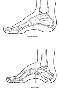
Cavus foot is a condition in which the foot has a very high arch. The high-arched foot places an excessive amount of weight on the ball and heel of the foot when walking or standing. Cavus foot can lead to a variety of signs and symptoms, such as pain and instability. It can develop at any age and can occur in one or both feet.
Causes of Cavus Foot (High-Arched Foot)
Cavus foot is often caused by a neurologic disorder or other medical condition, such as cerebral palsy, Charcot-Marie-Tooth disease, spina bifida, polio, muscular dystrophy or stroke. In other cases of cavus foot, the high arch may represent an inherited structural abnormality. An accurate diagnosis is important because the underlying cause of cavus foot largely determines its future course. If the high arch is due to a neurologic disorder or other medical condition, it is likely to progressively worsen. On the other hand, cases of cavus foot that do not result from neurologic disorders usually do not change in appearance.
Symptoms of Cavus Foot (High-Arched Foot)
The arch of a cavus foot will appear high even when standing. In addition, one or more of the following symptoms may be present:
- Hammertoes (bent toes) or claw toes (toes clenched like a fist)
- Calluses on the ball, side or heel of the foot
- Pain when standing or walking
- An unstable foot due to the heel tilting inward, which can lead to ankle sprains
Some people with cavus foot may also experience foot drop, a weakness of the muscles in the foot and ankle that results in dragging the foot when taking a step. Foot drop is usually a sign of an underlying neurologic condition.
Diagnosis of Cavus Foot (High-Arched Foot)
Diagnosis of cavus foot includes a review of the patient’s family history. The foot and ankle surgeon examines the foot, looking for a high arch and possible calluses, hammertoes and claw toes. The foot is tested for muscle strength, and the patient’s walking pattern and coordination are observed. If a neurologic condition appears to be present, the entire limb may be examined. The surgeon may also study the pattern of wear on the patient’s shoes.
X-rays are sometimes ordered to further assess the condition. In addition, the surgeon may refer the patient to a neurologist for a complete neurologic evaluation.
Nonsurgical Treatment
Nonsurgical treatment of cavus foot may include one or more of the following options:
- Orthotic devices. Custom orthotic devices that fit into the shoe can be beneficial because they provide stability and cushioning to the foot.
- Shoe modifications. High-topped shoes support the ankle, and shoes with heels a little wider on the bottom add stability.
- Bracing. The surgeon may recommend a brace to help keep the foot and ankle stable. Bracing is also useful in managing foot drop.
When Is Surgery Needed?
If nonsurgical treatment fails to adequately relieve pain and improve stability, surgery may be needed to decrease pain, increase stability and compensate for weakness in the foot. The surgeon will choose the best surgical procedure or combination of procedures based on the patient’s individual case. In some cases where an underlying neurologic problem exists, surgery may be needed again in the future due to the progression of the disorder.
Why choose a foot and ankle surgeon?
Foot and ankle surgeons are the leading experts in foot and ankle care today. As doctors of podiatric medicine – also known as podiatrists, DPMs or occasionally “foot and ankle doctors” – they are the board-certified surgical specialists of the podiatric profession. Foot and ankle surgeons have more education and training specific to the foot and ankle than any other healthcare provider.
Foot and ankle surgeons treat all conditions affecting the foot and ankle, from the simple to the complex, in patients of all ages including high-arched or Cavus foot. Their intensive education and training qualify foot and ankle surgeons to perform a wide range of surgeries, including any surgery that may be indicated for Cavus foot.
If you trim your toenails too short, particularly on the sides of your big toes, you may set the stage for an ingrown toenail. Like many people, when you trim your toenails, you may taper the corners so that the nail curves with the shape of your toe. But this technique may encourage your toenail to grow into the skin of your toe. The sides of the nail curl down and dig into your skin. An ingrown toenail may also happen if you wear shoes that are too tight or too short.
Symptoms
When you first have an ingrown toenail, it may be hard, swollen and tender. Later, it may get red and infected, and feel very sore. Ingrown toenails are a common, painful condition—particularly among teenagers. Any of your toenails can become ingrown, but the problem more often affects the big toe. An ingrown nail occurs when the skin on one or both sides of a nail grows over the edges of the nail, or when the nail itself grows into the skin. Redness, pain and swelling at the corner of the nail may result and infection may soon follow. Sometimes a small amount of pus can be seen draining from the area.
Ingrown nails may develop for many reasons. Some cases are congenital—the nail is just too large for the toe. Trauma, such as stubbing the toe or having the toe stepped on, may also cause an ingrown nail. However, the most common cause is tight shoe wear or improper grooming and trimming of the nail.
Treatment
Nonsurgical Treatment
Ingrown toenails should be treated as soon as they are recognized. If they are recognized early (before infection sets in), home care may prevent the need for further treatment:
- Soak the foot in warm water 3-4 times daily.
- Keep the foot dry during the rest of the day.
- Wear comfortable shoes with adequate room for the toes. Consider wearing sandals until the condition clears up.
- You may take ibuprofen or acetaminophen for pain relief.
- If there is no improvement in 2-3 days, or if the condition worsens, call your doctor.
You may need to gently lift the edge of the ingrown toenail from its embedded position and insert some cotton or waxed dental floss between the nail and your skin. Change this packing every day.
Surgical Treatment
If excessive inflammation, swelling, pain and discharge are present, the toenail is probably infected and should be treated by a physician (see left image below). You may need to take oral antibiotics and the nail may need to be partially or completely removed (see middle image below). The doctor can surgically remove a portion of the nail, a portion of the underlying nail bed, some of the adjacent soft tissues and even a part of the growth center (see right image below).
Surgery is effective in eliminating the nail edge from growing inward and cutting into the fleshy folds as the toenail grows forward. Permanent removal of the nail may be advised for children with chronic, recurrent infected ingrown toenails.
If you are in a lot of pain and/or the infection keeps coming back, your doctor may remove part of your ingrown toenail (partial nail avulsion). Your toe is injected with an anesthetic and your doctor uses scissors to cut away the ingrown part of the toenail, taking care not to disturb the nail bed. An exposed nail bed may be very painful. Removing your whole ingrown toenail (complete nail plate avulsion) increases the likelihood your toenail will come back deformed. It may take 3-4 months for your nail to regrow.
Risk Factors
Unless the problem is congenital, the best way to prevent ingrown toenails is to protect the feet from trauma and to wear shoes and hosiery (socks) with adequate room for the toes. Nails should be cut straight across with a clean, sharp nail trimmer without tapering or rounding the corners. Trim the nails no shorter than the edge of the toe. Keep the feet clean and dry at all times.
Intoeing, also known as pigeon-toed, is a condition in which the feet point inward when walking or standing. Outtoeing, on the other hand, is a condition in which the feet point outward. Both of these conditions can be caused by various factors, including muscle imbalances, bone abnormalities, or developmental problems.
Intoeing and outtoeing are most commonly seen in children and may improve on their own as the child grows and develops. However, if the condition persists or causes discomfort or difficulty walking, treatment may be necessary. Treatment options may include:
- Orthotic inserts to help correct the position of the feet
- Physical therapy to improve range of motion and strength in the feet and legs
- Surgery to correct underlying bone abnormalities
If your child is experiencing discomfort or difficulty walking due to intoeing or outtoeing, it is important to speak with a healthcare provider for proper diagnosis and treatment. In most cases, early treatment can help to alleviate discomfort and improve function.
Fractures of the Fifth Metatarsal
What Is a Fifth Metatarsal Fracture?
Fifth metatarsal fractures (breaks) are common foot foot injuries. The fifth metatarsal is the long bone on the outside of the foot that connects to the little toe. Two types of fractures that often occur in the fifth metatarsal are:

- Avulsion fracture. In an avulsion fracture, a small piece of bone is pulled off the main portion of the bone by a tendon or ligament. This type of fracture is the result of an injury in which the ankle rolls. Avulsion fractures are often overlooked when they occur with an ankle sprain.
- Jones fracture. Jones fractures occur in a small area of the fifth metatarsal that receives less blood and is therefore more prone to difficulties in healing. A Jones fracture can be either a stress fracture (a tiny hairline break that occurs over time) or an acute (sudden) break. Jones fractures are caused by overuse, repetitive stress or trauma. They are less common and more difficult to treat than avulsion fractures. Other types of fractures can occur in the fifth metatarsal. Examples include midshaft fractures, which usually result from trauma or twisting, and fractures of the metatarsal head and neck.
Symptoms of a Fifth Metatarsal Fracture
Avulsion and Jones fractures have the same signs and symptoms. These include:
- Pain, swelling and tenderness on the outside of the foot
- Difficulty walking
- Bruising
Diagnosis of a Fifth Metatarsal Fracture
Anyone who has symptoms of a fifth metatarsal fracture should see a foot and ankle surgeon as soon as possible for proper diagnosis and treatment. To arrive at a diagnosis, the surgeon will ask how the injury occurred or when the pain started. The foot will be examined, with the doctor gently pressing on different areas of the foot to determine where there is pain. The surgeon will also order x-rays. Because a Jones fracture sometimes does not show up on initial x-rays, additional imaging studies may be needed.
Nonsurgical Treatment
Until you are able to see a foot and ankle surgeon, the RICE method of care should be performed:
- Rest: Stay off the injured foot. Walking may cause further injury.
- Ice: Apply an ice pack to the injured area, placing a thin towel between the ice and the skin. Use ice for 20 minutes and then wait at least 40 minutes before icing again.
- Compression: An elastic wrap should be used to control swelling.
- Elevation: The foot should be raised slightly above the level of your heart to reduce swelling.
The foot and ankle surgeon may use one of these nonsurgical options for treatment of a fifth metatarsal fracture:
- Immobilization. Depending on the severity of the injury, the foot is kept immobile with a cast, cast boot or stiff-soled shoe. Crutches may also be needed to avoid placing weight on the injured foot.
- Bone stimulation. A pain-free external device is used to speed the healing of some fractures. Bone stimulation, most commonly used for Jones fractures, may be used as part of the treatment or following an inadequate response to immobilization.
When Is Surgery Needed?
If the injury involves a displaced bone, multiple breaks or has failed to adequately heal, surgery may be required. The foot and ankle surgeon will determine the type of procedure that is best suited to the individual patient.
Lisfranc (midfoot) injuries result if bones in the midfoot are broken or ligaments that support the midfoot are torn. The severity of a Lisfranc injury can vary widely — from a simple injury involving one midfoot joint to a complex injury involving many midfoot joints and broken bones.
A simple Lisfranc injury can be easily mistaken for a sprain, especially if the injury is a result of a straightforward twist and fall. However, injury to the Lisfranc joint is not a simple sprain that should be “walked off” or expected to heal quickly. Even a simple Lisfranc injury is a severe injury that may take many months to heal and may require surgery to treat.
Symptoms
These are the most common symptoms of Lisfranc injury:
- The top of foot may be swollen and painful.
- There may be bruising on both the top and bottom of the foot. Bruising on the bottom of the foot is highly suggestive of a Lisfranc injury.
- There may be pain in the midfoot that worsens with standing, walking, or attempting to push off on the affected foot. The pain can be so severe that weightbearing is not possible, and crutches may be required.
Treatment
Treatment for a Lisfranc injury depends on the severity of the injury.
Nonsurgical Treatment
If there are no fractures or dislocations in the joint and the ligaments are not completely torn, nonsurgical treatment may be all that is necessary for healing. A nonsurgical treatment plan includes wearing a non-weightbearing cast or boot for 6 to 8 weeks. You must be very strict about not putting weight on your injured foot during this period. This then progresses to weightbearing in a removable cast boot or an orthotic.
Surgical Treatment
Surgery is recommended for injuries with displaced fractures (broken bones) or with abnormal positioning (instability) of the joints. The goal of surgical treatment is to realign the joints, return the fractured bone fragments to a normal position, and restore stability to the midfoot.
Recovery
The goal in every patient is to have a pain-free, stable midfoot and return to all pre-injury activities. This goal can be achieved with either ORIF or fusion surgery in all types of patients. However, it is important to note that:
- The overall recovery process for resuming all activities may take 6 months to 1 year in some patients.
- Some patients may have persistent midfoot pain even with a successful surgery.
- Some athletic patients never return to their pre-injury levels of sport after these dificult injuries.
- Despite excellent surgical reduction and fixation, arthritis may occur from the damage to the cartilage. This may result in chronic pain and may require additional surgery in the future.
Midfoot arthritis is a type of osteoarthritis that affects the joints in the middle of the foot. It is a progressive condition that causes the cartilage in the joints to wear away, leading to pain, stiffness, and difficulty moving the foot. Midfoot arthritis is most commonly seen in people over the age of 50, but it can occur at any age.
Symptoms of midfoot arthritis may include:
- Pain and stiffness in the midfoot, especially when walking or standing
- Difficulty moving the foot or toes
- Swelling in the midfoot
- A feeling of instability in the foot
Midfoot arthritis is usually caused by wear and tear on the joints, but it can also be caused by injury or other conditions, such as rheumatoid arthritis.
Treatment for midfoot arthritis may include:
- Non-surgical options such as rest, ice, and over-the-counter pain medications
- Physical therapy to improve range of motion and strengthen the muscles around the joints
- Custom orthotic inserts to help redistribute pressure on the foot
- Surgery to fuse the joints or to remove damaged bone and cartilage
If you are experiencing pain or stiffness in your midfoot, it is important to speak with a healthcare provider for proper diagnosis and treatment. Early treatment can help to prevent the condition from becoming more severe and can help to alleviate discomfort and improve function.
A neuroma is an enlarged, benign growth of nerves, most commonly between the third and fourth toes. Neuromas are caused by tissue rubbing against and irritating the nerves. Pressure from poorly fitting shoes or an abnormal bone structure can also lead to this condition. Symptoms may include sensations of thickness, burning, numbness, tingling, or pain in the ball of the foot. Treatments generally include wearing corrective shoes or orthotics and/or receiving cortisone injections. In severe cases, surgical removal of the growth may be necessary.
Morton’s neuroma is a thickening of tissues around the nerve that leads to the toes. Morton’s neuroma usually develops between the third and fourth toes in response to irritation, such as that caused by wearing high-heeled or narrow shoes, or from trauma. Symptoms may include a burning pain that radiates from the ball of the foot to the toes or numbness in the toes. Conservative treatments usually resolve the pain or progressions of the condition, and range from wearing roomier, lower-heeled footwear or using orthotics to reduce the pressure on the nerve, to injections of corticosteroid medication to reduce swelling and inflammation.
A navicular stress fracture is a small crack or break in the navicular bone, which is a bone in the midfoot. Stress fractures are caused by repetitive stress or overuse, and they are most commonly seen in people who participate in high-impact activities, such as running or jumping.
Symptoms of a navicular stress fracture may include:
- Pain in the midfoot, especially when walking or standing
- Swelling in the midfoot
- Tenderness to touch in the midfoot
- Difficulty bearing weight on the affected foot
Treatment for a navicular stress fracture may include:
- Rest and ice to reduce swelling and inflammation
- Physical therapy to improve range of motion and strength in the foot and ankle
- Custom orthotic inserts to help redistribute pressure on the foot
- Medications to reduce pain and inflammation
- In severe cases, surgery may be necessary to repair the fracture
Os Trigonum Syndrome
What Is the Os Trigonum?
The os trigonum is an extra (accessory) bone that sometimes develops behind the ankle bone (talus). It is connected to the talus by a fibrous band. The presence of an os trigonum in one or both feet is congenital (present at birth). It becomes evident during adolescence when one area of the talus does not fuse with the rest of the bone, creating a small extra bone. Only a small number of people have this extra bone.
What Is Os Trigonum Syndrome?
Often, people do not know they have an os trigonum if it has not caused any problems. However, some people with this extra bone develop a painful condition known as os trigonum syndrome.
Os trigonum syndrome is usually triggered by an injury, such as an ankle sprain. The syndrome is also frequently caused by repeated downward pointing of the toes, which is common among ballet dancers, soccer players and other athletes.
For the person who has an os trigonum, pointing the toes downward can result in a “nutcracker injury.” Like an almond in a nutcracker, the os trigonum is crunched between the ankle and heel bones. As the os trigonum pulls loose, the tissue connecting it to the talus is stretched or torn and the area becomes inflamed.
Signs & Symptoms of Os Trigonum Syndrome
The signs and symptoms of os trigonum syndrome may include:
- Deep, aching pain in the back of the ankle, occurring mostly when pushing off on the big toe (as in walking) or when pointing the toes downward
- Tenderness in the area when touched
- Swelling in the back of the ankle
Diagnosis
Os trigonum syndrome can mimic other conditions, such as an Achilles tendon injury, ankle sprain or talus fracture. Diagnosis of os trigonum syndrome begins with questions from the doctor about the development of symptoms. After the foot and ankle are examined, x-rays or other imaging tests are often ordered to assist in making the diagnosis.
Treatment: Nonsurgical Approaches
Relief of the symptoms is often achieved through treatments that can include a combination of the following:
- Rest. It is important to stay off the injured foot to let the inflammation subside.
- Immobilization. A walking boot is often used to restrict ankle motion and to allow the injured tissue to heal.
- Ice. Swelling is decreased by applying a bag of ice covered with a thin towel to the affected area. Do not put ice directly against the skin.
- Oral medication. Nonsteroidal anti-inflammatory drugs (NSAIDs), such as ibuprofen, may be helpful in reducing the pain and inflammation.
- Injections. Sometimes cortisone is injected into the area to reduce the inflammation and pain.
When Is Surgery Needed?
Most patients’ symptoms improve with nonsurgical treatment. However, in some patients, surgery may be required to relieve the symptoms. Surgery typically involves removal of the os trigonum, as this extra bone is not necessary for normal foot function.
Talar Dome Lesion
What Is a Talar Dome Lesion? 
The ankle joint is composed of the bottom of the tibia (shin) bone and the top of the talus (ankle) bone. The top of the talus is dome-shaped and is completely covered with cartilage—a tough, rubbery tissue that enables the ankle to move smoothly. A talar dome lesion is an injury to the cartilage and underlying bone of the talus within the ankle joint. It is also called an osteochondral defect (OCD) or osteochondral lesion of the talus (OLT). “Osteo” means bone and “chondral” refers to cartilage.
Talar dome lesions are usually caused by an injury, such as an ankle sprain. If the cartilage does not heal properly following the injury, it softens and begins to break off. Sometimes a broken piece of the damaged cartilage and bone will float in the ankle.
Signs & Symptoms
Unless the injury is extensive, it may take months, a year or even longer for symptoms to develop. The signs and symptoms of a talar dome lesion may include:
- Chronic pain deep in the ankle—typically worse when bearing weight on the foot (especially during sports) and less when resting
- An occasional clicking or catching feeling in the ankle when walking
- A sensation of the ankle locking or giving out
- Episodes of swelling of the ankle—occurring when bearing weight and subsiding when at rest
Diagnosis
A talar dome lesion can be difficult to diagnose because the precise site of the pain can be hard to pinpoint. To diagnose this injury, the foot and ankle surgeon will question the patient about recent or previous injury and will examine the foot and ankle, moving the ankle joint to help determine if there is pain, clicking or limited motion within that joint.
Sometimes the surgeon will inject the joint with an anesthetic (pain-relieving medication) to see if the pain goes away for a while, indicating that the pain is coming from inside the joint. X-rays are taken, and often an MRI or other advanced imaging tests are ordered to further evaluate the lesion and extent of the injury.
Nonsurgical Treatment Approaches
Treatment depends on the severity of the talar dome lesion. If the lesion is stable (without loose pieces of cartilage or bone), one or more of the following nonsurgical treatment options may be considered:
- Immobilization. Depending on the type of injury, the leg may be placed in a cast or cast boot to protect the talus. During this period of immobilization, nonweightbearing range-of-motion exercises may be recommended.
- Oral medications. Nonsteroidal anti-inflammatory drugs (NSAIDs), such as ibuprofen, may be helpful in reducing the pain and inflammation.
- Physical therapy. Range-of-motion and strengthening exercises are beneficial once the lesion is adequately healed. Physical therapy may also include techniques to reduce pain and swelling.
- Ankle brace. Wearing an ankle brace may help protect the patient from reinjury if the ankle is unstable.
When Is Surgery Needed?
If nonsurgical treatment fails to relieve the symptoms of talar dome lesions, surgery may be necessary. Surgery may involve removal of the loose bone and cartilage fragments within the joint and establishing an environment for healing. A variety of surgical techniques is available to accomplish this. The surgeon will select the best procedure based on the specific case.
Complications of Talar Dome Lesions
Depending on the amount of damage to the cartilage in the ankle joint, arthritis may develop in the joint, resulting in chronic pain, swelling and limited joint motion. Treatment for these complications is best directed by a foot and ankle surgeon and may include one or more of the following:
- Nonsteroidal or steroidal anti-inflammatory medications
- Physical therapy
- Bracing
- Surgical intervention
Peroneal tendinosis is a condition that affects the peroneal tendons, which are the tendons that run along the outside of the ankle and foot. The condition is caused by overuse or repetitive stress on the tendons, leading to inflammation and degeneration. Peroneal tendinosis is often seen in people who participate in high-impact activities, such as running or jumping.
Symptoms of peroneal tendinosis may include:
- Pain and tenderness along the outside of the ankle and foot
- Swelling in the ankle and foot
- Weakness in the foot and ankle
- Difficulty bearing weight on the affected leg
Treatment for peroneal tendinosis may include:
- Rest and ice to reduce swelling and inflammation
- Stretching and strengthening exercises to improve flexibility and support the foot
- Custom orthotic inserts to help redistribute pressure on the foot
- Physical therapy to improve range of motion and strength in the foot and ankle
- Medications to reduce pain and inflammation
- In severe cases, surgery may be necessary to repair damaged tendons
A pilon fracture is a type of break that occurs at the bottom of the tibia (shinbone) and involves the weight-bearing surface of the ankle joint. With this type of injury, the other bone in the lower leg, the fibula, is frequently broken as well. A pilon fracture typically occurs as the result of a high-energy event, such as a car collision or fall from a height. Pilon fractures are often severe injuries that can permanently affect the ankle joint.
In most cases, surgery is needed to restore the damaged bone to its normal position. Because of the energy required to cause a pilon fracture, patients may have other injuries that require treatment as well.
Symptoms
Patients with pilon fractures usually experience immediate and severe pain. Other symptoms may include:
- Swelling
- Bruising
- Tenderness
- Inability to bear weight on the injured leg
- Deformity — your ankle may look angled or crooked
Treatment
Many pilon fractures require surgery but, rarely, some stable fractures can be treated nonsurgically. Whether or not your doctor recommends surgery often depends on how out of place (displaced) the fractured pieces of bone are.
Plantar Fasciitis (Heel Pain)
Heel pain is most often caused by plantar fasciitis, a condition that is sometimes also called heel spur syndrome when a spur is present. Heel pain may also be due to other causes such as a stress fracture, tendonitis, arthritis, nerve irritation or, rarely, a cyst. Because there are several potential causes, it is important to have heel pain properly diagnosed.
What Is Plantar Fasciitis?
Plantar fasciitis is an inflammation of the band of tissue (the plantar fascia) that extends from the heel to the toes. In this condition, the fascia first becomes irritated and then inflamed, resulting in heel pain.
Causes
The most common cause of plantar fasciitis relates to faulty structure of the foot. For example, people who have
problems with their arches, either overly flat feet or high-arched feet are more prone to developing plantar fasciitis. Wearing non-supportive footwear on hard, flat surfaces puts abnormal strain on the plantar fascia and can also lead to plantar fasciitis. This is particularly evident when one’s job requires long hours on the feet. Obesity may also contribute to plantar fasciitis.
Symptoms
The symptoms of plantar fasciitis are:
• Pain on the bottom of the heel
• Pain the is usually worse upon arising
• Pain that increases over several months
People with plantar fasciitis often describe the pain as being worse when they get up in the morning or after they’ve been sitting for long periods of time. The pain decreases after a few minutes of walking because walking stretches the fascia. For some people the pain subsides but returns after spending long periods of time on their feet.
Diagnosis
To arrive at a diagnosis, your foot and ankle specialist will obtain your medical history and examine your foot. Throughout
this process, your doctor rules out all the possible causes for your heel pain other than plantar fasciitis. In
addition, diagnostic imaging studies such as x-rays or other imaging modalities may be used to distinguish the
different types of heel pain. Sometimes heel spurs are found in patients with plantar fasciitis, but the spurs are rarely a source of pain. When they are present, the condition may be diagnosed as plantar fasciitis/heel spur syndrome.
Non-Surgical Treatment
Treatment of plantar fasciitis begins with first-line strategies which can be initiated at home:
• Stretching exercises. Exercises that stretch out the calf muscles and plantar fascia help ease pain and assist with recovery.
• Avoid going barefoot. When you walk without shoes, you put undue strain and stress on your plantar fascia.
• Ice. Putting an ice pack on your heel for 20 minutes several times a day helps reduce inflammation. Place a thin towel between the ice and your heel and never apply ice directly to the skin.
• Limit activities. Cut down on extended physical activities to give your heel a rest.
• Shoe modifications. Wearing supportive shoes that have a good arch support and a slightly raised heel reduces stress on the plantar fascia.
• Medications. Oral nonsteroidal anti-inflammatory drugs (NSAIDs) such as ibuprofen may be recommended to reduce pain and inflammation.
• Padding and strapping. Placing pads in the shoe softens the impact of walking. Strapping helps support the foot and reduce strain on the fascia.
If you still have pain after several weeks, your foot doctor may add one or more of these treatment approaches:
• Orthotic devices. Custom orthotic devices that fit into your shoe help correct the underlying structural abnormalities causing the plantar fasciitis.
• Injection therapy. In some cases, corticosteroid injections are used to help reduce the inflammation and relieve pain.
• Removable walking cast. A removable walking cast may be used to keep your foot immobile for a few weeks to allow it to rest and heal.
• Night splint. Wearing a night splint allows you to maintain an extended stretch of the plantar fascia while sleeping. This may help reduce the morning pain experienced by some patients.
• Physical therapy. Exercises and other physical therapy measures may be used to help provide relief.
When Is Surgery Needed?
Although most patients with plantar fasciitis respond to non-surgical treatment, a small percentage of patients may require surgery. If the pain persists after several months of non-surgical treatment, surgery can be considered. Your foot and ankle surgeon will discuss the surgical options with you and determine which approach would be most beneficial for you.
Long Term Care
No matter what kind of treatment you undergo for plantar fasciitis, the underlying causes that led to this condition may remain. Therefore, you will need to continue with preventive measures. Wearing supportive shoes, stretching, and using custom orthotic devices are the mainstay of long-term treatment success against plantar fasciitis.
Plantar Fibroma
What Is the Plantar Fibroma?
A plantar fibroma is a fibrous knot (nodule) in the arch of the foot. It is embedded within the plantar fascia, a band of tissue that extends from the heel to the toes on the bottom of the foot. A plantar fibroma can develop in one or both feet, is benign (nonmalignant) and usually will not go away or get smaller without treatment. Definitive causes for this condition have not been clearly identified, but there are several options for the treatment of plantar fibroma.


Signs & Symptoms of Plantar Fibroma
The characteristic sign of a plantar fibroma is a noticeable lump in the arch that feels firm to the touch. This mass can remain the same size or get larger over time or additional fibromas may develop.
People who have a plantar fibroma may or may not have pain. When pain occurs, it is often caused by shoes pushing against the lump in the arch, although it can also arise when walking or standing barefoot.
Diagnosis of Plantar Fibroma
To diagnose a plantar fibroma, the foot and ankle surgeon will examine the foot and press on the affected area. Sometimes this can produce pain that extends down to the toes. An MRI or biopsy may be performed to further evaluate the lump and aid in diagnosis.
Treatment of Plantar Fibroma
Nonsurgical treatment may help relieve the pain of a plantar fibroma, although it will not make the mass disappear. The foot and ankle surgeon may select one or more of the following nonsurgical options:
- Steroid injections. Injecting corticosteroid medication into the mass may help shrink it and thereby relieve the pain that occurs when walking. This reduction may only be temporary and the fibroma could slowly return to its original size.
- Orthotic devices. If the fibroma is stable, meaning it is not changing in size, custom orthotic devices (shoe inserts) may relieve the pain by distributing the patient’s weight away from the fibroma.
- Physical therapy. The pain is sometimes treated through physical therapy methods that deliver anti-inflammatory medication into the fibroma without the need for injection.
If the mass increases in size or pain, the patient should be further evaluated.
Surgical treatment to remove the fibroma is considered if the patient continues to experience pain following nonsurgical approaches. Surgical removal of a plantar fibroma may result in a flattening of the arch or development of hammertoes. Orthotic devices may be prescribed to provide support to the foot. Due to the high incidence of recurrence with this condition, continued follow-up with the foot and ankle surgeon is recommended.
Why choose a foot and ankle surgeon?
Foot and ankle surgeons are the leading experts in foot and ankle care today. As doctors of podiatric medicine – also known as podiatrists, DPMs or occasionally “foot and ankle doctors” – they are the board-certified surgical specialists of the podiatric profession. Foot and ankle surgeons have more education and training specific to the foot and ankle than any other healthcare provider.
Foot and ankle surgeons treat all conditions affecting the foot and ankle, from the simple to the complex, in patients of all ages including plantar fibromas. Their intensive education and training qualify foot and ankle surgeons to perform a wide range of surgeries, including any surgery that may be indicated for plantar fibromas.
Warts are caused by a virus that lives within the deepest layer of the skin, making them difficult to treat. Those that appear on the sole of the foot are called plantar warts. Plantar warts are usually benign. Children, especially teenagers, tend to be more susceptible to warts than adults are. Warts are usually treated conservatively, but can be very stubborn to completely resolve. Plantar warts tend to be hard and flat, with a rough surface and well-defined boundaries; warts that appear on the top of the foot or on the toes are generally raised and fleshier. Sometimes, warts can be confused for corns or rough skin. It is important to note that warts can be very resistant to treatment and have a tendency to re-occur.
The plantar wart is often contracted by walking barefoot on dirty surfaces or littered ground where the virus is lurking. The causative virus thrives in warm, moist environments, making infection a common occurrence in communal bathing facilities.
SYMPTOMS
Most warts are harmless, but may be painful. Plantar warts are often gray or brown (but the color may vary), with a center that appears as one or more pinpoints of black. When plantar warts develop on the weight-bearing areas of the foot (the ball of the foot, or the heel, for example), they can be the source of sharp, burning pain.
TREATMENT
First-line therapy for warts in our office is trimming down the dead skin on top of and surrounding the wart. This is usually painless and allows for medication to penetrate the lesion more effectively. Next, a medication called Cantharone is applied directly to the skin. Cantharone comes from a bug called a blister beetle that kills other insects by spraying a substance that causes blistering. This medication does not hurt upon initial application, but may sting 2-3 days later. After two weeks, the patient returns and the blistered wart is painlessly trimmed away. Sometimes additional treatments may be needed. Our physician has been using this medication with great success.
For more stubborn warts, a simple surgical procedure to excise the wart, performed under local anesthetic, may be indicated. A procedure known as CO2 laser cautery is performed under local anesthesia in an outpatient surgery facility to excise the lesion and cauterize bleeding tissue. The laser reduces post-treatment scarring and is a safe form for eliminating wart lesions. Postoperative care involves daily foot soaks for a week and daily bandaid applications.
Posterior Tibial Tendon Dysfunction (PTTD)
What Is PTTD? 
The posterior tibial tendon serves as one of the major supporting structures of the foot, helping it to function while walking. Posterior tibial tendon dysfunction (PTTD) is a condition caused by changes in the tendon, impairing its ability to support the arch. This results in flattening of the foot.
PTTD is often called adult acquired flatfoot because it is the most common type of flatfoot developed during adulthood. Although this condition typically occurs in only one foot, some people may develop it in both feet. PTTD is usually progressive, which means it will keep getting worse, especially if it is not treated early.
Causes
Overuse of the posterior tibial tendon is often the cause of PTTD. In fact, the symptoms usually occur after activities that involve the tendon, such as running, walking, hiking or climbing stairs.
Symptoms
The symptoms of PTTD may include pain, swelling, a flattening of the arch and an inward rolling of the ankle. As the condition progresses, the symptoms will change.
For example, when PTTD initially develops, there is pain on the inside of the foot and ankle (along the course of the tendon). In addition, the area may be red, warm and swollen.
Later, as the arch begins to flatten, there may still be pain on the inside of the foot and ankle. But at this point, the foot and toes begin to turn outward and the ankle rolls inward.
As PTTD becomes more advanced, the arch flattens even more and the pain often shifts to the outside of the foot, below the ankle. The tendon has deteriorated considerably, and arthritis often develops in the foot. In more severe cases, arthritis may also develop in the ankle.
Nonsurgical Treatment
Because of the progressive nature of PTTD, early treatment is advised. If treated early enough, your symptoms may resolve without the need for surgery, and progression of your condition can be arrested.
In contrast, untreated PTTD could leave you with an extremely flat foot, painful arthritis in the foot and ankle and increasing limitations on walking, running or other activities.
In many cases of PTTD, treatment can begin with nonsurgical approaches that may include:
- Orthotic devices or bracing. To give your arch the support it needs, your foot and ankle surgeon may provide you with an ankle brace or a custom orthotic device that fits into the shoe.
- Immobilization. Sometimes a short-leg cast or boot is worn to immobilize the foot and allow the tendon to heal, or you may need to completely avoid all weightbearing for a while.
- Physical therapy. Ultrasound therapy and exercises may help rehabilitate the tendon and muscle following immobilization.
- Medications. Nonsteroidal anti-inflammatory drugs (NSAIDs), such as ibuprofen, help reduce the pain and inflammation.
- Shoe modifications. Your foot and ankle surgeon may advise changes to your shoes and may provide special inserts designed to improve arch support.
When Is Surgery Needed?
In cases of PTTD that have progressed substantially or have failed to improve with nonsurgical treatment, surgery may be required. For some advanced cases, surgery may be the only option. Your foot and ankle surgeon will determine the best approach for you.
Rheumatoid Arthritis in the Foot and Ankle
What Is Rheumatoid Arthritis?
Rheumatoid arthritis (RA) is a disease in which certain cells of the immune system malfunction and attack healthy joints.
RA causes inflammation in the lining (synovium) of joints, most often the joints of the hands and feet. The signs of inflammation can include pain, swelling, redness and a feeling of warmth around affected joints. In some patients, chronic inflammation results in damage to the cartilage and bones in the joint. Serious damage can lead to permanent joint destruction, deformity and disability.
When joints become inflamed due to RA, the synovium thickens and produces an excess of joint fluid. This overabundance of fluid, along with inflammatory chemicals released by the immune system, causes swelling and damage to the joint’s cartilage and bones.
Symptoms Affecting the Foot & Ankle
Foot problems caused by RA commonly occur in the forefoot (the ball of the foot, near the toes), although RA can also affect other areas of the foot and ankle. The most common signs and symptoms of RA-related foot problems, in addition to the abnormal appearance of deformities, are pain, swelling, joint stiffness and difficulty walking.
Deformities and conditions associated with RA may include:
- Rheumatoid nodules (lumps), which cause pain when they rub against shoes or, if they appear on the bottom of the foot, pain when walking
- Dislocated toe joints
- Hammertoes
- Bunions
- Heel pain
- Achilles tendon pain
- Flatfoot ankle pain
Diagnosis
RA is diagnosed on the basis of a clinical examination as well as blood tests.
To further evaluate the patient’s foot and ankle problems, the surgeon may order x-rays and/or other imaging tests.
Treatment by the Foot & Ankle Surgeon
While treatment of RA focuses on the medication prescribed by a patient’s primary doctor or rheumatologist, the foot and ankle surgeon will develop a treatment plan aimed at relieving the pain of RA-related foot problems. The plan may include one or more of the following options:
- Orthotic devices. The surgeon often fits the patient with custom orthotic devices to provide cushioning for rheumatoid nodules, minimize pain when walking and give needed support to improve the foot’s mechanics.
- Accommodative shoes. These are used to relieve pressure and pain and to assist with walking.
- Aspiration of fluid. When inflammation flares up in a joint, the surgeon may aspirate (draw out) fluid to reduce the swelling and pain.
- Steroid injections. Injections of anti-inflammatory medication may be applied directly to an inflamed joint or to a rheumatoid nodule.
When Is Surgery Needed?
When RA produces pain and deformity in the foot that is not relieved through other treatments, surgery may be required. The foot and ankle surgeon will select the procedure best suited to the patient’s condition and lifestyle.
Sesamoid Injuries in the Foot
What are the Sesamoids of the Foot? 
To understand sesamoid injuries in the foot, it’s imporant to know what sesamoinds are. A sesamoid is a bone embedded in a tendon. Sesamoids are found in several joints in the body. In the normal foot, the sesamoids are two pea-shaped bones located in the ball of the foot, beneath the big toe joint.
Acting as a pulley for tendons, the sesamoids help the big toe move normally and provide leverage when the big toe pushes off during walking and running. The sesamoids also serve as a weightbearing surface for the first metatarsal bone (the long bone connected to the big toe), absorbing the weight placed on the ball of the foot when walking, running and jumping.
Sesamoid injuries can involve the bones, tendons and/or surrounding tissue in the joint. They are often associated with activities requiring increased pressure on the ball of the foot, such as running, basketball, football, golf, tennis and ballet. In addition, people with high arches are at risk for developing sesamoid problems. Frequent wearing of high-heeled shoes can also be a contributing factor.
Types of Sesamoid Injuries in the Foot
There are three types of sesamoid injuries in the foot:
- Turf toe. This is an injury of the soft tissue surrounding the big toe joint. It usually occurs when the big toe joint is extended beyond its normal range. Turf toe causes immediate, sharp pain and swelling. It usually affects the entire big toe joint and limits the motion of the toe. Turf toe may result in an injury to the soft tissue attached to the sesamoid or a fracture of the sesamoid. Sometimes a “pop” is felt at the moment of injury.
- Fracture. A fracture (break) in a sesamoid bone can be either acute or chronic.

- An acute fracture is caused by trauma—a direct blow or impact to the bone. An acute sesamoid fracture produces immediate pain and swelling at the site of the break but usually does not affect the entire big toe joint. A chronic fracture is a stress fracture (a hairline break usually caused by repetitive stress or overuse).
- A chronic sesamoid fracture produces longstanding pain in the ball of the foot beneath the big toe joint. The pain, which tends to come and go, generally is aggravated with activity and relieved with rest.
- Sesamoiditis. This is an overuse injury involving chronic inflammation of the sesamoid bones and the tendons involved with those bones. Sesamoiditis is caused by increased pressure on the sesamoids. Often, sesamoiditis is associated with a dull, longstanding pain beneath the big toe joint. The pain comes and goes, usually occurring with certain shoes or certain activities.
Diagnosis of Sesamoids Injuries in the Foot
In diagnosing a sesamoid injury, the foot and ankle surgeon will examine the foot, focusing on the big toe joint. The surgeon will press on the big toe, move it up and down, and may assess the patient’s walking and evaluate the wear pattern on the patient’s shoes. X-rays are ordered, and in some cases, advanced imaging studies may be ordered.
Nonsurgical Treatment
Nonsurgical treatment for sesamoid injuries of the foot may include one or more of the following options, depending on the type of injury and degree of severity:
- Padding, strapping or taping. A pad may be placed in the shoe to cushion the inflamed sesamoid area, or the toe may be taped or strapped to relieve that area of tension.
- Immobilization. The foot may be placed in a cast or removable walking cast. Crutches may be used to prevent placing weight on the foot.
- Oral medications. Nonsteroidal anti-inflammatory drugs (NSAIDs), such as ibuprofen, are often helpful in reducing the pain and inflammation.
- Physical therapy. The rehabilitation period following immobilization sometimes includes physical therapy, such as exercises (range of motion, strengthening and conditioning) and ultrasound therapy.
- Steroid injections. In some cases, cortisone is injected into the joint to reduce pain and inflammation.
- Orthotic devices. Custom orthotic devices that fit into the shoe may be prescribed for long-term treatment of sesamoiditis to balance the pressure placed on the ball of the foot.
When Is Surgery Needed?
When sesamoid injuries fail to respond to nonsurgical treatment, surgery may be required. The foot and ankle surgeon will determine the type of procedure that is best suited to the individual patient
Sinus tarsi syndrome is a condition that affects the sinus tarsi, which is a small, fluid-filled space located near the ankle. It is a condition that can cause pain and instability in the ankle, especially when walking or standing on uneven surfaces. Sinus tarsi syndrome is often caused by injury or overuse of the ankle, but it can also be caused by underlying conditions, such as arthritis or flat feet.
Symptoms of sinus tarsi syndrome may include:
- Pain and swelling on the outside of the ankle
- Instability in the ankle, especially when walking or standing on uneven surfaces
- Difficulty bearing weight on the affected leg
- Numbness or tingling in the foot
Treatment for sinus tarsi syndrome may include:
- Rest and ice to reduce swelling and inflammation
- Stretching and strengthening exercises to improve flexibility and support the ankle
- Physical therapy to improve range of motion and strength in the foot and ankle
- Custom orthotic inserts to help redistribute pressure on the foot
- Medications to reduce pain and inflammation
- In severe cases, surgery may be necessary to repair damaged tissue or stabilize the ankle
Stress Fracture in the Foot
Stress fractures are tiny hairline breaks that can occur in the bones of the foot. They can be caused by overtraining or overuse, improper training habits or surfaces, improper shoes, flatfoot or other foot deformities and osteoporosis. These tiny breaks in the bones of the feet can lead to a complete break if left untreated.
Pain, swelling, redness and bruising can be signs of a stress fracture. The fracture can occur almost anywhere in the foot. X-rays and other studies are used to diagnose the stress fracture. A foot and ankle surgeon should be seen as early as possible to start treatment and possibly shorten the recovery time. Possible treatments include rest and possible immobilization of the foot. In some cases, surgery may be required to stabilize the stress fracture or to repair a stress fracture that has progressed to a fracture.
A talus fracture is a break in the talus bone, which is a small bone in the ankle that connects the leg bones (tibia and fibula) to the foot bones (calcaneus and navicular). Talus fractures can be caused by a variety of factors, including falls, motor vehicle accidents, and sports injuries.
Symptoms of a talus fracture may include:
- Pain and swelling in the ankle
- Difficulty bearing weight on the affected leg
- Bruising or discoloration in the ankle
- Difficulty moving the foot or ankle
Treatment for a talus fracture may include:
- Rest and ice to reduce swelling and inflammation
- Compression bandages to support the injury and help control swelling
- Physical therapy to improve range of motion and strength in the foot and ankle
- Medications to reduce pain and inflammation
- In severe cases, surgery may be necessary to repair the fracture and stabilize the ankle
A tarsal coalition is an abnormal connection of two or more bones in the foot. The bones affected — called tarsal bones — are located toward the back of the foot and in the heel, and the connection of the bones can result in a severe, rigid flatfoot.
Although tarsal coalition is often present at birth, children typically do not show signs of the disorder until early adolescence. The foot may become stiff and painful, and everyday physical activities are often difficult.
For many children with tarsal coalition, symptoms are relieved with simple treatments, such as orthotics and physical therapy. If a child has severe symptoms that do not respond to simple treatments and continue to interfere with their daily activities, surgery may be recommended.
Symptoms
Many tarsal coalitions are never discovered because they do not cause symptoms or any obvious foot deformity. When symptoms do occur, they may include:
- Stiff, painful feet. The pain usually occurs below the ankle around the middle or back half of the foot.
- A rigid, flat foot that makes it difficult to walk on uneven surfaces. To accommodate for the foot’s lack of motion, the patient may roll the ankle more than normal, which may result in recurrent ankle sprains.
- Increased pain or a limp with higher levels of activity.
Treatment
Tarsal coalitions only require treatment if they are causing symptoms.
Nonsurgical Treatment
- Rest. Taking a break from high-impact activity for a period time — 3 to 6 weeks — can reduce stress on the tarsal bones and relieve pain.
- Orthotics. Arch supports, shoe inserts like heel cups and wedges, and other types of orthotics may be recommended to help stabilize the foot and relieve pain.
- Temporary boot or cast. These options can immobilize the foot and take stress off of the tarsal bones.
- Injections. Steroid medications may be used in conjunction with other nonsurgical options to provide temporary pain relief.
Surgical Treatment
When nonsurgical treatments are not effective at easing pain or improving function, your doctor may consider surgery.
The surgical procedure your doctor recommends will depend on the size and location of the coalition, as well as whether the joints between the bones show signs of arthritis.
Recovery
Depending upon the type and location of your surgery, a cast will be required for a period of time to protect the surgical site and prevent you from putting weight on the foot. Casts are typically replaced with walking boots, and your doctor may recommend physical therapy exercises to begin restoring range of motion and strength.
Your doctor will determine when it is safe for you to begin putting weight on your foot. Arch supports or orthotics may also be helpful in stabilizing the joint, even after surgery.
Although it may take several months to fully recover, most patients have pain relief and improved motion after surgery.
Tarsal tunnel syndrome is a condition where the tibial nerve (the largest nerve to enter the foot) is compressed, causing pain and numbness. The compression of the nerve occurs at the tarsal tunnel, a canal formed at the inner part of the ankle, just behind the medial malleolus, that large bony bump on the inside part of the ankle.
CAUSES
Tarsal tunnel syndrome can be caused by injury, disease, or due to the natural shaping of the foot. Causes include:
SYMPTOMS
. Pain and tingling around the ankle
- Swelling in the foot
Pain radiating up into the leg or down into the ankle and foot with a pins and needles feeling in the foot.
TREATMENT :
Tarsal tunnel syndrome can be treated conservatively; however, surgical intervention may be warranted. The goal of conservative therapy is to reduce the swelling in the ankle that causes compression of the nerve.
Conservative Options
Anti-Inflammatory Medication—Early treatment may involve oral or injectable anti-inflammatory medication to reduce the swelling in the inner ankle area
Rest and Icing—Used to relieve pain and reduce swelling
Bracing and Strapping—May be used for correcting biomechanical imbalance, controlling excessive pronation, and supporting the ligaments and tendons attached to the heel bone. This helps reduce motion at the ankle that can cause compression of the nerve.
Custom-Molded Orthotics—These custom-made shoe inserts are useful in controlling foot function and to relieve pressure from the inner ankle.
Surgical Treatment
For more serious cases of tarsal tunnel syndrome, surgical intervention to reduce the underlying cause of the compression may be needed. In such cases the surgeon would decompress the nerve and relieve it of any impingements that may cause compression.
Since fungal nails are usually more resistant and more difficult to treat , topical or oral antifungal medications may be prescribed. Permanent nail removal is another possible form of treatment for fungal nails.
After a fungal nail infection has cleared up, you can take steps to prevent the infection from coming back.
Keeping the fungus under control will help prevent a fungal infection of the skin from reinfecting the nail. Before bed, thoroughly wash and dry your feet, and apply a non-prescription anti-fungal cream to the entire foot from the ankle down. Use the cream every night, then gradually apply it less often. Keep your feet dry. Dry feet are less likely to become infected. Apply powder to your dry feet after you take a shower or bath.
Other tips:
- Don’t share nail clippers or nail files with others.
- Don’t share shoes or socks with others.
- Try not to injure your nail, such as by cutting it too short (trauma to the nail may lead to infections).
- Wear dry cotton socks, and change them two or three times a day if necessary.
- Wear dry shoes that allow air to circulate around your feet (tight, enclosed, moist shoes contribute to fungal toenail infections).
- Wear shower sandals or shower shoes when you are at a public pool or shower.
Prevention
Follow basic foot care guidelines and you more than likely can head off most common foot fungus problems.
The simplest definition of turf toe is that it is a sprain of the main joint of the big toe. The injury happens when the toe is forcibly bent up into hyperextension, such as when pushing off into a sprint and having the toe get stuck flat on the ground.
First Aid Treatment
The RICE protocol is effective for most sports-related injuries when they first occur. RICE stands for:
- Rest. Take a break from the activity that caused the injury and avoid walking or putting weight on your foot.
- Ice. Use cold packs for 20 minutes at a time, several times a day. Do not apply ice directly to the skin.
- Compression. To help prevent additional swelling, wear an elastic compression bandage.
- Elevation. To reduce swelling, recline when you rest, and prop your leg up so it is higher than your heart.
In addition, over-the-counter anti-inflammatory medications, such as ibuprofen and naproxen, can help provide symptom relief.
Treatment
Nonsurgical Treatment
Most cases of turf toe are treated without surgery. Nonsurgical treatment is determined by the grade of the injury.
Surgical Treatment
Surgery is often not necessary for treating turf toe. However, if your symptoms persist or your level of athletic play is affected, surgery may be an option. Doctors most often recommend surgery for larger Grade 3 injuries, such as:
- A severe tear of the plantar complex
- Fracture of the sesamoid
- Vertical instability (unusual up and down motion) of the MTP joint
- Loose bony chip in the joint
- Damage to the cartilage of the joint
- New or worsening bunion (turning outward of the big toe)
The surgical procedure will vary according to the injury. The aim of surgery is to repair the soft tissues and restore the MTP joint motion to preserve normal function.
Long-Term Outcomes
Turf toe injuries that are addressed early typically heal fairly well.
A wide range of mild to moderate, but persistent symptoms — such as pain and joint stiffness — are the most common complications.
Potential long-term complications include lack of push-off strength, stiffness, bunion, and cocking up of the big toe.
Wounds
Ulcers, which are open sores in the skin, occur when the outer layers of the skin are injured and the deeper tissues become exposed. They can be caused by excess pressure due to ill-fitting shoes, long periods in bed or after an injury that breaks the skin. Ulcers are commonly seen in patients living with diabetes, neuropathy or vascular disease. Open wounds can put patients at increased risk of developing infection in the skin and bone.
The signs and symptoms of ulcers may include drainage, odor or red, inflamed, thickened tissue. Pain may or may not be present.
Diagnosis may include x-rays to evaluate possible bone involvement. Other advanced imaging studies may also be ordered to evaluate for vascular disease, which may affect a patient’s ability to heal the wound.
Ulcers are treated by removing the unhealthy tissue and performing local wound care to assist in healing. Special shoes or padding may be used to remove excess pressure on the area. If infection is present, antibiotics will be necessary. In severe cases that involve extensive infection or are slow to heal, surgery or other advanced wound care treatments may be necessary.